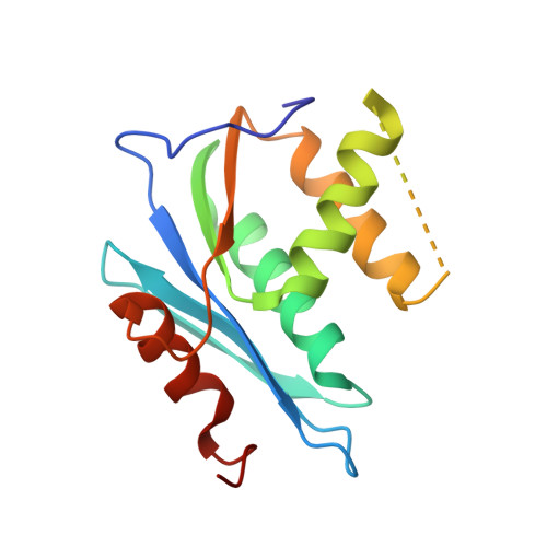
3QIN

Crystal Structure of HIV-1 RNase H p15 with engineered E. coli loop and pyrimidinol carboxylic acid inhibitor


Experimental Data Snapshot

  • Method: X-RAY DIFFRACTION
  • Resolution: 1.70 Å
  • R-Value Free: 0.253 
  • R-Value Work: 0.220 
  • R-Value Observed: 0.223 

wwPDB Validation   3D Report Full Report


Ligand Structure Quality Assessment 


This is version 1.5 of the entry. See complete history


Literature

Structural and Binding Analysis of Pyrimidinol Carboxylic Acid and N-Hydroxy Quinazolinedione HIV-1 RNase H Inhibitors.

Lansdon, E.B.Liu, Q.Leavitt, S.A.Balakrishnan, M.Perry, J.K.Lancaster-Moyer, C.Kutty, N.Liu, X.Squires, N.H.Watkins, W.J.Kirschberg, T.A.

(2011) Antimicrob Agents Chemother 55: 2905-2915

  • DOI: https://doi.org/10.1128/AAC.01594-10
  • Primary Citation of Related Structures:  
    3QIN, 3QIO, 3QIP

  • PubMed Abstract: 

    HIV-1 RNase H breaks down the intermediate RNA-DNA hybrids during reverse transcription, requiring two divalent metal ions for activity. Pyrimidinol carboxylic acid and N-hydroxy quinazolinedione inhibitors were designed to coordinate the two metal ions in the active site of RNase H. High-resolution (1.4 Å to 2.1 Å) crystal structures were determined with the isolated RNase H domain and reverse transcriptase (RT), which permit accurate assessment of the metal and water environment at the active site. The geometry of the metal coordination suggests that the inhibitors mimic a substrate state prior to phosphodiester catalysis. Surface plasmon resonance studies confirm metal-dependent binding to RNase H and demonstrate that the inhibitors do not bind at the polymerase active site of RT. Additional evaluation of the RNase H site reveals an open protein surface with few additional interactions to optimize active-site inhibitors.


  • Organizational Affiliation

    Gilead Sciences, Inc., 333 Lakeside Dr., Foster City, CA 94404, USA. eric.lansdon@gilead.com


Macromolecules
Find similar proteins by:  (by identity cutoff)  |  3D Structure
Entity ID: 1
MoleculeChains Sequence LengthOrganismDetailsImage
Fusion protein of HIV-1 RNase H p15 with engineered E. coli loop150HIV-1 M:B_HXB2REscherichia coli K-12Mutation(s): 0 
Gene Names: gag-polrnhAdasFherArnhsdrAb0214JW0204
EC: 3.4.23.16 (PDB Primary Data), 2.7.7.49 (PDB Primary Data), 2.7.7.7 (PDB Primary Data), 3.1.26.13 (PDB Primary Data), 3.1.13.2 (PDB Primary Data), 2.7.7 (PDB Primary Data), 3.1 (PDB Primary Data), 3.1.26.4 (PDB Primary Data)
UniProt
Find proteins for P0A7Y4 (Escherichia coli (strain K12))
Explore P0A7Y4 
Go to UniProtKB:  P0A7Y4
Find proteins for P04585 (Human immunodeficiency virus type 1 group M subtype B (isolate HXB2))
Explore P04585 
Go to UniProtKB:  P04585
Entity Groups  
Sequence Clusters30% Identity50% Identity70% Identity90% Identity95% Identity100% Identity
UniProt GroupsP04585P0A7Y4
Sequence Annotations
Expand
  • Reference Sequence
Small Molecules
Binding Affinity Annotations 
IDSourceBinding Affinity
P1Y PDBBind:  3QIN Kd: 136 (nM) from 1 assay(s)
Binding MOAD:  3QIN Kd: 136 (nM) from 1 assay(s)
Experimental Data & Validation

Experimental Data

  • Method: X-RAY DIFFRACTION
  • Resolution: 1.70 Å
  • R-Value Free: 0.253 
  • R-Value Work: 0.220 
  • R-Value Observed: 0.223 
  • Space Group: I 21 21 21
Unit Cell:
Length ( Å )Angle ( ˚ )
a = 38.841α = 90
b = 90.37β = 90
c = 111.922γ = 90
Software Package:
Software NamePurpose
CrystalCleardata collection
PHASERphasing
PHENIXrefinement
HKL-2000data reduction
HKL-2000data scaling

Structure Validation

View Full Validation Report


Ligand Structure Quality Assessment 


Entry History 

Deposition Data

Revision History  (Full details and data files)

  • Version 1.0: 2011-04-20
    Type: Initial release
  • Version 1.1: 2011-07-13
    Changes: Version format compliance
  • Version 1.2: 2017-05-31
    Changes: Database references
  • Version 1.3: 2021-01-13
    Changes: Derived calculations, Structure summary
  • Version 1.4: 2021-11-17
    Changes: Database references, Structure summary
  • Version 1.5: 2023-09-13
    Changes: Data collection, Refinement description