3QIB

Crystal structure of the 2B4 TCR in complex with MCC/I-Ek


Experimental Data Snapshot

  • Method: X-RAY DIFFRACTION
  • Resolution: 2.70 Å
  • R-Value Free: 0.246 
  • R-Value Work: 0.221 
  • R-Value Observed: 0.222 

wwPDB Validation   3D Report Full Report


Ligand Structure Quality Assessment 


This is version 1.3 of the entry. See complete history


Literature

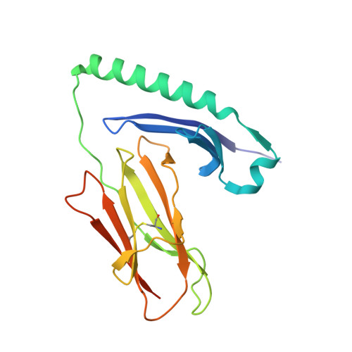
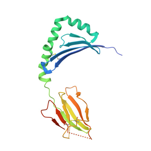
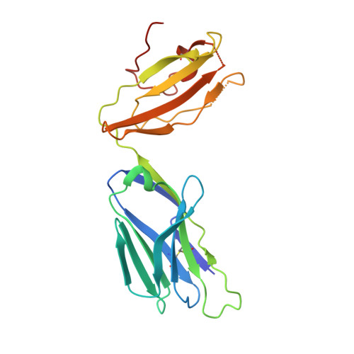
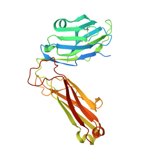
Structural basis of specificity and cross-reactivity in T cell receptors specific for cytochrome c-I-E(k).

Newell, E.W.Ely, L.K.Kruse, A.C.Reay, P.A.Rodriguez, S.N.Lin, A.E.Kuhns, M.S.Garcia, K.C.Davis, M.M.

(2011) J Immunol 186: 5823-5832

  • DOI: https://doi.org/10.4049/jimmunol.1100197
  • Primary Citation of Related Structures:  
    3QIB, 3QIU, 3QIW, 3QJF, 3QJH

  • PubMed Abstract: 

    T cells specific for the cytochrome c Ag are widely used to investigate many aspects of TCR specificity and interactions with peptide-MHC, but structural information has long been elusive. In this study, we present structures for the well-studied 2B4 TCR, as well as a naturally occurring variant of the 5c.c7 TCR, 226, which is cross-reactive with more than half of possible substitutions at all three TCR-sensitive residues on the peptide Ag. These structures alone and in complex with peptide-MHC ligands allow us to reassess many prior mutagenesis results. In addition, the structure of 226 bound to one peptide variant, p5E, shows major changes in the CDR3 contacts compared with wild-type, yet the TCR V-region contacts with MHC are conserved. These and other data illustrate the ability of TCRs to accommodate large variations in CDR3 structure and peptide contacts within the constraints of highly conserved TCR-MHC interactions.


  • Organizational Affiliation

    Department of Microbiology and Immunology, Stanford University School of Medicine, Stanford University, Stanford, CA 94305, USA.


Macromolecules
Find similar proteins by:  (by identity cutoff)  |  3D Structure
Entity ID: 1
MoleculeChains Sequence LengthOrganismDetailsImage
H-2 class II histocompatibility antigen, E-K alpha chain193Mus musculusMutation(s): 0 
UniProt
Find proteins for P04224 (Mus musculus)
Explore P04224 
Go to UniProtKB:  P04224
Entity Groups  
Sequence Clusters30% Identity50% Identity70% Identity90% Identity95% Identity100% Identity
UniProt GroupP04224
Sequence Annotations
Expand
  • Reference Sequence
Find similar proteins by:  (by identity cutoff)  |  3D Structure
Entity ID: 2
MoleculeChains Sequence LengthOrganismDetailsImage
MHC class II E-beta-k202Mus musculusMutation(s): 0 
Gene Names: H2-Eb1
UniProt & NIH Common Fund Data Resources
Find proteins for P04230 (Mus musculus)
Explore P04230 
Go to UniProtKB:  P04230
IMPC:  MGI:95901
Entity Groups  
Sequence Clusters30% Identity50% Identity70% Identity90% Identity95% Identity100% Identity
UniProt GroupP04230
Sequence Annotations
Expand
  • Reference Sequence
Find similar proteins by:  (by identity cutoff)  |  3D Structure
Entity ID: 3
MoleculeChains Sequence LengthOrganismDetailsImage
T-cell receptor alpha chain C region207Homo sapiensMutation(s): 0 
Gene Names: TCRATRAC
UniProt & NIH Common Fund Data Resources
Find proteins for P01848 (Homo sapiens)
Explore P01848 
Go to UniProtKB:  P01848
PHAROS:  P01848
Entity Groups  
Sequence Clusters30% Identity50% Identity70% Identity90% Identity95% Identity100% Identity
UniProt GroupP01848
Sequence Annotations
Expand
  • Reference Sequence
Find similar proteins by:  (by identity cutoff)  |  3D Structure
Entity ID: 4
MoleculeChains Sequence LengthOrganismDetailsImage
2B4 beta chain270Mus musculusMutation(s): 0 
Entity Groups  
Sequence Clusters30% Identity50% Identity70% Identity90% Identity95% Identity100% Identity
Sequence Annotations
Expand
  • Reference Sequence

Find similar proteins by:  Sequence   |   3D Structure  

Entity ID: 5
MoleculeChains Sequence LengthOrganismDetailsImage
MCC peptideE [auth P]13Manduca sextaMutation(s): 0 
UniProt
Find proteins for P00039 (Manduca sexta)
Explore P00039 
Go to UniProtKB:  P00039
Entity Groups  
Sequence Clusters30% Identity50% Identity70% Identity90% Identity95% Identity100% Identity
UniProt GroupP00039
Sequence Annotations
Expand
  • Reference Sequence
Small Molecules
Ligands 4 Unique
IDChains Name / Formula / InChI Key2D Diagram3D Interactions
NAG
Query on NAG

Download Ideal Coordinates CCD File 
F [auth B],
I [auth C]
2-acetamido-2-deoxy-beta-D-glucopyranose
C8 H15 N O6
OVRNDRQMDRJTHS-FMDGEEDCSA-N
BMA
Query on BMA

Download Ideal Coordinates CCD File 
K [auth C]beta-D-mannopyranose
C6 H12 O6
WQZGKKKJIJFFOK-RWOPYEJCSA-N
FUC
Query on FUC

Download Ideal Coordinates CCD File 
G [auth B],
J [auth C]
alpha-L-fucopyranose
C6 H12 O5
SHZGCJCMOBCMKK-SXUWKVJYSA-N
PEG
Query on PEG

Download Ideal Coordinates CCD File 
H [auth B]
L [auth C]
M [auth C]
N [auth C]
O [auth D]
H [auth B],
L [auth C],
M [auth C],
N [auth C],
O [auth D],
P [auth D],
Q [auth D]
DI(HYDROXYETHYL)ETHER
C4 H10 O3
MTHSVFCYNBDYFN-UHFFFAOYSA-N
Experimental Data & Validation

Experimental Data

  • Method: X-RAY DIFFRACTION
  • Resolution: 2.70 Å
  • R-Value Free: 0.246 
  • R-Value Work: 0.221 
  • R-Value Observed: 0.222 
  • Space Group: F 2 2 2
Unit Cell:
Length ( Å )Angle ( ˚ )
a = 146.749α = 90
b = 167.178β = 90
c = 259.489γ = 90
Software Package:
Software NamePurpose
PHENIXrefinement

Structure Validation

View Full Validation Report


Ligand Structure Quality Assessment 


Entry History 

Deposition Data

Revision History  (Full details and data files)

  • Version 1.0: 2011-04-27
    Type: Initial release
  • Version 1.1: 2011-07-13
    Changes: Version format compliance
  • Version 1.2: 2011-08-10
    Changes: Database references
  • Version 1.3: 2020-07-29
    Type: Remediation
    Reason: Carbohydrate remediation
    Changes: Data collection, Database references, Derived calculations, Structure summary