3QI9

Crystal structure of mouse CD1d-alpha-phosphotidylinositol with mouse Valpha14-Vbeta6 2A3-D NKT TCR


Experimental Data Snapshot

  • Method: X-RAY DIFFRACTION
  • Resolution: 2.30 Å
  • R-Value Free: 0.275 
  • R-Value Work: 0.247 
  • R-Value Observed: 0.248 

wwPDB Validation   3D Report Full Report


Ligand Structure Quality Assessment 


This is version 2.1 of the entry. See complete history


Literature

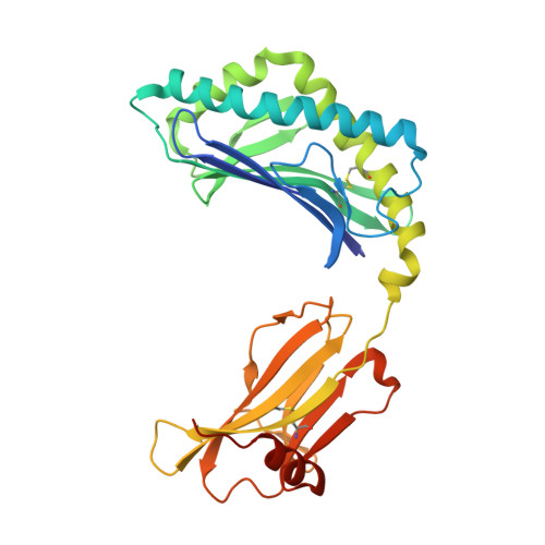
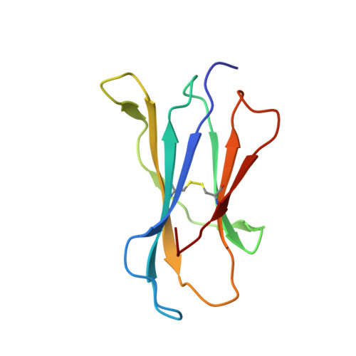
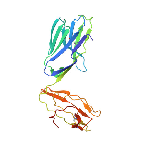
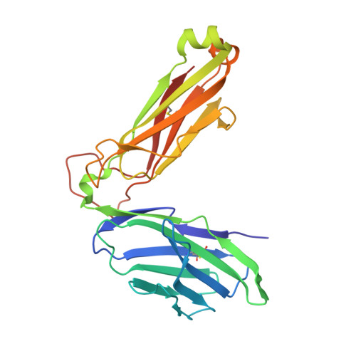
A molecular basis for NKT cell recognition of CD1d-self-antigen

Mallevaey, T.Clarke, A.J.Scott-Browne, J.P.Young, M.H.Roisman, L.C.Pellicci, D.G.Patel, O.Vivian, J.P.Matsuda, J.L.McCluskey, J.Godfrey, D.I.Marrack, P.Rossjohn, J.Gapin, L.

(2011) Immunity 34: 315-326

  • DOI: https://doi.org/10.1016/j.immuni.2011.01.013
  • Primary Citation of Related Structures:  
    3QI9

  • PubMed Abstract: 

    The antigen receptor for natural killer T cells (NKT TCR) binds CD1d-restricted microbial and self-lipid antigens, although the molecular basis of self-CD1d recognition is unclear. Here, we have characterized NKT TCR recognition of CD1d molecules loaded with natural self-antigens (Ags) and report the 2.3 Å resolution structure of an autoreactive NKT TCR-phosphatidylinositol-CD1d complex. NKT TCR recognition of self- and foreign antigens was underpinned by a similar mode of germline-encoded recognition of CD1d. However, NKT TCR autoreactivity is mediated by unique sequences within the non-germline-encoded CDR3β loop encoding for a hydrophobic motif that promotes self-association with CD1d. Accordingly, NKT cell autoreactivity may arise from the inherent affinity of the interaction between CD1d and the NKT TCR, resulting in the recognition of a broad range of CD1d-restricted self-antigens. This demonstrates that multiple self-antigens can be recognized in a similar manner by autoreactive NKT TCRs.


  • Organizational Affiliation

    Department of Immunology, University of Colorado School of Medicine and National Jewish Health, Denver, CO 80206, USA.


Macromolecules
Find similar proteins by:  (by identity cutoff)  |  3D Structure
Entity ID: 1
MoleculeChains Sequence LengthOrganismDetailsImage
Antigen-presenting glycoprotein CD1d1302Mus musculusMutation(s): 0 
Gene Names: Cd1d1Cd1.1
UniProt & NIH Common Fund Data Resources
Find proteins for P11609 (Mus musculus)
Explore P11609 
Go to UniProtKB:  P11609
IMPC:  MGI:107674
Entity Groups  
Sequence Clusters30% Identity50% Identity70% Identity90% Identity95% Identity100% Identity
UniProt GroupP11609
Sequence Annotations
Expand
  • Reference Sequence
Find similar proteins by:  (by identity cutoff)  |  3D Structure
Entity ID: 2
MoleculeChains Sequence LengthOrganismDetailsImage
Beta-2-microglobulin99Mus musculusMutation(s): 0 
Gene Names: B2m
UniProt & NIH Common Fund Data Resources
Find proteins for P01887 (Mus musculus)
Explore P01887 
Go to UniProtKB:  P01887
IMPC:  MGI:88127
Entity Groups  
Sequence Clusters30% Identity50% Identity70% Identity90% Identity95% Identity100% Identity
UniProt GroupP01887
Sequence Annotations
Expand
  • Reference Sequence
Find similar proteins by:  (by identity cutoff)  |  3D Structure
Entity ID: 3
MoleculeChains Sequence LengthOrganismDetailsImage
NKT TCR V alpha 14207Mus musculusMutation(s): 0 
Entity Groups  
Sequence Clusters30% Identity50% Identity70% Identity90% Identity95% Identity100% Identity
Sequence Annotations
Expand
  • Reference Sequence
Find similar proteins by:  (by identity cutoff)  |  3D Structure
Entity ID: 4
MoleculeChains Sequence LengthOrganismDetailsImage
NKT TCR V beta 6 2A3-D243Mus musculusMutation(s): 0 
Entity Groups  
Sequence Clusters30% Identity50% Identity70% Identity90% Identity95% Identity100% Identity
Sequence Annotations
Expand
  • Reference Sequence
Oligosaccharides

Help

Entity ID: 5
MoleculeChains Length2D Diagram Glycosylation3D Interactions
2-acetamido-2-deoxy-beta-D-glucopyranose-(1-4)-2-acetamido-2-deoxy-beta-D-glucopyranose
E
2N-Glycosylation
Glycosylation Resources
GlyTouCan:  G42666HT
GlyCosmos:  G42666HT
GlyGen:  G42666HT
Small Molecules
Ligands 4 Unique
IDChains Name / Formula / InChI Key2D Diagram3D Interactions
PII
Query on PII

Download Ideal Coordinates CCD File 
H [auth A]2-[(HYDROXY{[(2R,3R,5S,6R)-2,3,4,5,6-PENTAHYDROXYCYCLOHEXYL]OXY}PHOSPHORYL)OXY]-1-[(PALMITOYLOXY)METHYL]ETHYL HEPTADECANOATE
C42 H81 O13 P
QVAGJHIIXLLVCI-KSGPIMHQSA-N
NAG
Query on NAG

Download Ideal Coordinates CCD File 
F [auth A],
G [auth A]
2-acetamido-2-deoxy-beta-D-glucopyranose
C8 H15 N O6
OVRNDRQMDRJTHS-FMDGEEDCSA-N
GOL
Query on GOL

Download Ideal Coordinates CCD File 
J [auth B],
K [auth D]
GLYCEROL
C3 H8 O3
PEDCQBHIVMGVHV-UHFFFAOYSA-N
MG
Query on MG

Download Ideal Coordinates CCD File 
I [auth A]MAGNESIUM ION
Mg
JLVVSXFLKOJNIY-UHFFFAOYSA-N
Experimental Data & Validation

Experimental Data

  • Method: X-RAY DIFFRACTION
  • Resolution: 2.30 Å
  • R-Value Free: 0.275 
  • R-Value Work: 0.247 
  • R-Value Observed: 0.248 
  • Space Group: P 41 21 2
Unit Cell:
Length ( Å )Angle ( ˚ )
a = 95.697α = 90
b = 95.697β = 90
c = 289.71γ = 90
Software Package:
Software NamePurpose
REFMACrefinement
PDB_EXTRACTdata extraction
MOSFLMdata reduction
SCALAdata scaling
PHASERphasing

Structure Validation

View Full Validation Report


Ligand Structure Quality Assessment 


Entry History 

Deposition Data

Revision History  (Full details and data files)

  • Version 1.0: 2012-02-15
    Type: Initial release
  • Version 1.1: 2020-01-29
    Changes: Advisory, Database references, Derived calculations, Source and taxonomy
  • Version 2.0: 2020-07-29
    Type: Remediation
    Reason: Carbohydrate remediation
    Changes: Atomic model, Data collection, Derived calculations, Structure summary
  • Version 2.1: 2023-11-01
    Changes: Advisory, Data collection, Database references, Refinement description, Structure summary