3QDA

Crystal structure of W95L beta-2 microglobulin


Experimental Data Snapshot

  • Method: X-RAY DIFFRACTION
  • Resolution: 1.57 Å
  • R-Value Free: 0.198 
  • R-Value Work: 0.147 
  • R-Value Observed: 0.149 

wwPDB Validation   3D Report Full Report


This is version 1.2 of the entry. See complete history


Literature

The two tryptophans of beta2-microglobulin have distinct roles in function and folding and might represent two independent responses to evolutionary pressure.

Raimondi, S.Barbarini, N.Mangione, P.Esposito, G.Ricagno, S.Bolognesi, M.Zorzoli, I.Marchese, L.Soria, C.Bellazzi, R.Monti, M.Stoppini, M.Stefanelli, M.Magni, P.Bellotti, V.

(2011) BMC Evol Biol 11: 159-159

  • DOI: https://doi.org/10.1186/1471-2148-11-159
  • Primary Citation of Related Structures:  
    3QDA

  • PubMed Abstract: 

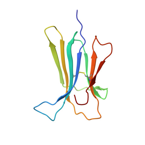
    We have recently discovered that the two tryptophans of human β2-microglobulin have distinctive roles within the structure and function of the protein. Deeply buried in the core, Trp95 is essential for folding stability, whereas Trp60, which is solvent-exposed, plays a crucial role in promoting the binding of β2-microglobulin to the heavy chain of the class I major histocompatibility complex (MHCI). We have previously shown that the thermodynamic disadvantage of having Trp60 exposed on the surface is counter-balanced by the perfect fit between it and a cavity within the MHCI heavy chain that contributes significantly to the functional stabilization of the MHCI. Therefore, based on the peculiar differences of the two tryptophans, we have analysed the evolution of β2-microglobulin with respect to these residues.


  • Organizational Affiliation

    Department of Biochemistry, University of Pavia, via Taramelli 3b, 27100 Pavia, Italy.


Macromolecules
Find similar proteins by:  (by identity cutoff)  |  3D Structure
Entity ID: 1
MoleculeChains Sequence LengthOrganismDetailsImage
Beta-2-microglobulin100Homo sapiensMutation(s): 1 
Gene Names: B2MCDABP0092HDCMA22PNM_004048
UniProt & NIH Common Fund Data Resources
Find proteins for P61769 (Homo sapiens)
Explore P61769 
Go to UniProtKB:  P61769
PHAROS:  P61769
GTEx:  ENSG00000166710 
Entity Groups  
Sequence Clusters30% Identity50% Identity70% Identity90% Identity95% Identity100% Identity
UniProt GroupP61769
Sequence Annotations
Expand
  • Reference Sequence
Small Molecules
Ligands 1 Unique
IDChains Name / Formula / InChI Key2D Diagram3D Interactions
PGE
Query on PGE

Download Ideal Coordinates CCD File 
B [auth A]TRIETHYLENE GLYCOL
C6 H14 O4
ZIBGPFATKBEMQZ-UHFFFAOYSA-N
Experimental Data & Validation

Experimental Data

  • Method: X-RAY DIFFRACTION
  • Resolution: 1.57 Å
  • R-Value Free: 0.198 
  • R-Value Work: 0.147 
  • R-Value Observed: 0.149 
  • Space Group: C 1 2 1
Unit Cell:
Length ( Å )Angle ( ˚ )
a = 77.41α = 90
b = 28.98β = 121.77
c = 54.45γ = 90
Software Package:
Software NamePurpose
MxCuBEdata collection
MOLREPphasing
REFMACrefinement
MOSFLMdata reduction
SCALAdata scaling

Structure Validation

View Full Validation Report


Entry History 

Deposition Data

Revision History  (Full details and data files)

  • Version 1.0: 2011-06-29
    Type: Initial release
  • Version 1.1: 2011-07-13
    Changes: Version format compliance
  • Version 1.2: 2023-09-13
    Changes: Data collection, Database references, Derived calculations, Refinement description