3PWR

HIV-1 Protease Mutant L76V complexed with Saquinavir


Experimental Data Snapshot

  • Method: X-RAY DIFFRACTION
  • Resolution: 1.45 Å
  • R-Value Free: 0.197 
  • R-Value Observed: 0.145 

wwPDB Validation   3D Report Full Report


Ligand Structure Quality Assessment 


This is version 1.4 of the entry. See complete history


Literature

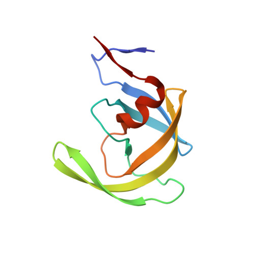
The L76V Drug Resistance Mutation Decreases the Dimer Stability and Rate of Autoprocessing of HIV-1 Protease by Reducing Internal Hydrophobic Contacts.

Louis, J.M.Zhang, Y.Sayer, J.M.Wang, Y.F.Harrison, R.W.Weber, I.T.

(2011) Biochemistry 50: 4786-4795

  • DOI: https://doi.org/10.1021/bi200033z
  • Primary Citation of Related Structures:  
    3PWM, 3PWR

  • PubMed Abstract: 

    The mature HIV-1 protease (PR) bearing the L76V drug resistance mutation (PR(L76V)) is significantly less stable, with a >7-fold higher dimer dissociation constant (K(d)) of 71 ± 24 nM and twice the sensitivity to urea denaturation (UC(50) = 0.85 M) relative to those of PR. Differential scanning calorimetry showed decreases in T(m) of 12 °C for PR(L76V) in the absence of inhibitors and 5-7 °C in the presence of inhibitors darunavir (DRV), saquinavir (SQV), and lopinavir (LPV), relative to that of PR. Isothermal titration calorimetry gave a ligand dissociation constant of 0.8 nM for DRV, ∼160-fold higher than that of PR, consistent with DRV resistance. Crystal structures of PR(L76V) in complexes with DRV and SQV were determined at resolutions of 1.45-1.46 Å. Compared to the corresponding PR complexes, the mutated Val76 lacks hydrophobic interactions with Asp30, Lys45, Ile47, and Thr74 and exhibits closer interactions with Val32 and Val56. The bound DRV lacks one hydrogen bond with the main chain of Asp30 in PR(L76V) relative to PR, possibly accounting for the resistance to DRV. SQV shows slightly improved polar interactions with PR(L76V) compared to those with PR. Although the L76V mutation significantly slows the N-terminal autoprocessing of the precursor TFR-PR(L76V) to give rise to the mature PR(L76V), the coselected M46I mutation counteracts the effect by enhancing this rate but renders the TFR-PR(M46I/L76V) precursor less responsive to inhibition by 6 μM LPV while preserving inhibition by SQV and DRV. The correlation of lowered stability, higher K(d), and impaired autoprocessing with reduced internal hydrophobic contacts suggests a novel molecular mechanism for drug resistance.


  • Organizational Affiliation

    Laboratory of Chemical Physics, National Institute of Diabetes and Digestive and Kidney Diseases, National Institutes of Health, Bethesda, MD 20892, USA. johnl@intra.niddk.nih.gov


Macromolecules
Find similar proteins by:  (by identity cutoff)  |  3D Structure
Entity ID: 1
MoleculeChains Sequence LengthOrganismDetailsImage
Protease
A, B
99Human immunodeficiency virus 1Mutation(s): 6 
Gene Names: pol
EC: 3.4.23.16
UniProt
Find proteins for P03367 (Human immunodeficiency virus type 1 group M subtype B (isolate BRU/LAI))
Explore P03367 
Go to UniProtKB:  P03367
Entity Groups  
Sequence Clusters30% Identity50% Identity70% Identity90% Identity95% Identity100% Identity
UniProt GroupP03367
Sequence Annotations
Expand
  • Reference Sequence
Small Molecules
Ligands 3 Unique
IDChains Name / Formula / InChI Key2D Diagram3D Interactions
ROC
Query on ROC

Download Ideal Coordinates CCD File 
C [auth A](2S)-N-[(2S,3R)-4-[(2S,3S,4aS,8aS)-3-(tert-butylcarbamoyl)-3,4,4a,5,6,7,8,8a-octahydro-1H-isoquinolin-2-yl]-3-hydroxy-1 -phenyl-butan-2-yl]-2-(quinolin-2-ylcarbonylamino)butanediamide
C38 H50 N6 O5
QWAXKHKRTORLEM-UGJKXSETSA-N
GOL
Query on GOL

Download Ideal Coordinates CCD File 
E [auth A]
F [auth A]
G [auth A]
H [auth A]
I [auth A]
E [auth A],
F [auth A],
G [auth A],
H [auth A],
I [auth A],
J [auth A],
K [auth B],
L [auth B]
GLYCEROL
C3 H8 O3
PEDCQBHIVMGVHV-UHFFFAOYSA-N
CL
Query on CL

Download Ideal Coordinates CCD File 
D [auth A]CHLORIDE ION
Cl
VEXZGXHMUGYJMC-UHFFFAOYSA-M
Binding Affinity Annotations 
IDSourceBinding Affinity
ROC BindingDB:  3PWR Ki: min: 0.04, max: 138 (nM) from 25 assay(s)
Kd: min: 0.31, max: 67 (nM) from 6 assay(s)
IC50: min: 0.2, max: 270 (nM) from 16 assay(s)
-TΔS: min: -8.03e+1, max: -2.80e+1 (kJ/mol) from 45 assay(s)
ΔH: min: -3.18e+1, max: 43.47 (kJ/mol) from 45 assay(s)
ΔG: min: -6.23e+1, max: -3.55e+1 (kJ/mol) from 43 assay(s)
Biologically Interesting Molecules (External Reference) 1 Unique
Experimental Data & Validation

Experimental Data

  • Method: X-RAY DIFFRACTION
  • Resolution: 1.45 Å
  • R-Value Free: 0.197 
  • R-Value Observed: 0.145 
  • Space Group: P 21 21 2
Unit Cell:
Length ( Å )Angle ( ˚ )
a = 58.84α = 90
b = 86.17β = 90
c = 46.24γ = 90
Software Package:
Software NamePurpose
MAR345data collection
MOLREPphasing
SHELXL-97refinement
HKL-2000data reduction
HKL-2000data scaling

Structure Validation

View Full Validation Report


Ligand Structure Quality Assessment 


Entry History 

Deposition Data

Revision History  (Full details and data files)

  • Version 1.0: 2011-04-20
    Type: Initial release
  • Version 1.1: 2011-07-13
    Changes: Version format compliance
  • Version 1.2: 2012-12-12
    Changes: Other
  • Version 1.3: 2017-11-08
    Changes: Refinement description
  • Version 1.4: 2023-09-13
    Changes: Data collection, Database references, Derived calculations, Refinement description, Structure summary