3PMO

The structure of LpxD from Pseudomonas aeruginosa at 1.3 A resolution


Experimental Data Snapshot

  • Method: X-RAY DIFFRACTION
  • Resolution: 1.30 Å
  • R-Value Free: 0.185 
  • R-Value Work: 0.164 
  • R-Value Observed: 0.165 

wwPDB Validation   3D Report Full Report


This is version 1.2 of the entry. See complete history


Literature

The structure of LpxD from Pseudomonas aeruginosa at 1.3 A resolution.

Badger, J.Chie-Leon, B.Logan, C.Sridhar, V.Sankaran, B.Zwart, P.H.Nienaber, V.

(2011) Acta Crystallogr Sect F Struct Biol Cryst Commun 67: 749-752

  • DOI: https://doi.org/10.1107/S1744309111018811
  • Primary Citation of Related Structures:  
    3PMO

  • PubMed Abstract: 

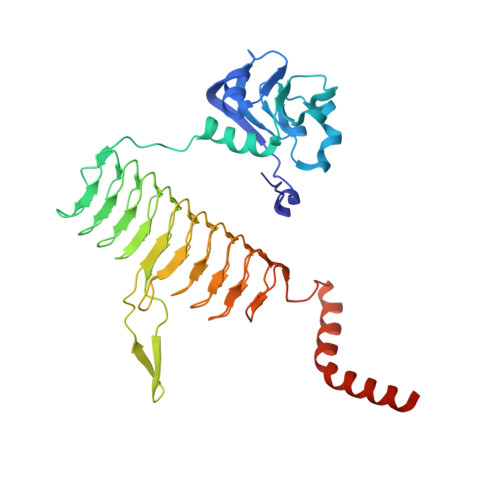
    LpxD is a bacterial protein that is part of the biosynthesis pathway of lipid A and is responsible for transferring 3-hydroxymyristic acid from the R-3-hydroxymyristoyl-acyl carrier protein to the 2-OH group of UDP-3-O-(3-hydroxymyristoyl) glucosamine. The crystal structure of LpxD from Pseudomonas aeruginosa has been determined at high resolution (1.3 Å). The crystal belonged to space group H3, with unit-cell parameters a=b=106.19, c=93.38 Å, and contained one molecule in the asymmetric unit. The structure was solved by molecular replacement using the known structure of LpxD from Escherichia coli (PDB entry 3eh0) as a search model and was refined to Rwork=16.4% (Rfree=18.5%) using 91,655 reflections. The final protein model includes 355 amino-acid residues (including 16 amino acids from a 20 amino-acid N-terminal His tag), one chloride ion and two ethylene glycol molecules.


  • Organizational Affiliation

    Zenobia Therapeutics, 505 Coast Boulevard South, Suite 111, La Jolla, CA 92122, USA. john@zenobiatherapeutics.com


Macromolecules
Find similar proteins by:  (by identity cutoff)  |  3D Structure
Entity ID: 1
MoleculeChains Sequence LengthOrganismDetailsImage
UDP-3-O-[3-hydroxymyristoyl] glucosamine N-acyltransferase372Pseudomonas aeruginosaMutation(s): 0 
Gene Names: lpxDPA3646
EC: 2.3.1
UniProt
Find proteins for Q9HXY6 (Pseudomonas aeruginosa (strain ATCC 15692 / DSM 22644 / CIP 104116 / JCM 14847 / LMG 12228 / 1C / PRS 101 / PAO1))
Explore Q9HXY6 
Go to UniProtKB:  Q9HXY6
Entity Groups  
Sequence Clusters30% Identity50% Identity70% Identity90% Identity95% Identity100% Identity
UniProt GroupQ9HXY6
Sequence Annotations
Expand
  • Reference Sequence
Experimental Data & Validation

Experimental Data

  • Method: X-RAY DIFFRACTION
  • Resolution: 1.30 Å
  • R-Value Free: 0.185 
  • R-Value Work: 0.164 
  • R-Value Observed: 0.165 
  • Space Group: H 3
Unit Cell:
Length ( Å )Angle ( ˚ )
a = 106.19α = 90
b = 106.19β = 90
c = 93.38γ = 120
Software Package:
Software NamePurpose
ADSCdata collection
MOLREPphasing
REFMACrefinement
MOSFLMdata reduction
SCALAdata scaling

Structure Validation

View Full Validation Report


Entry History 

Deposition Data

Revision History  (Full details and data files)

  • Version 1.0: 2011-07-27
    Type: Initial release
  • Version 1.1: 2012-03-28
    Changes: Database references
  • Version 1.2: 2024-02-21
    Changes: Data collection, Database references, Derived calculations