3PB1

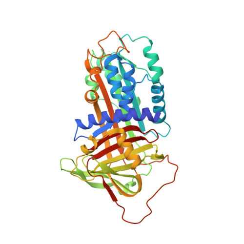
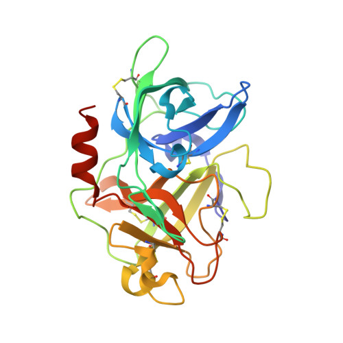
Crystal Structure of a Michaelis Complex between Plasminogen Activator Inhibitor-1 and Urokinase-type Plasminogen Activator


Experimental Data Snapshot

  • Method: X-RAY DIFFRACTION
  • Resolution: 2.30 Å
  • R-Value Free: 0.272 
  • R-Value Work: 0.220 
  • R-Value Observed: 0.223 

wwPDB Validation   3D Report Full Report


This is version 1.2 of the entry. See complete history


Literature

Structural basis for recognition of urokinase-type plasminogen activator by plasminogen activator inhibitor-1.

Lin, Z.Jiang, L.Yuan, C.Jensen, J.K.Zhang, X.Luo, Z.Furie, B.C.Furie, B.Andreasen, P.A.Huang, M.

(2011) J Biol Chem 286: 7027-7032

  • DOI: https://doi.org/10.1074/jbc.M110.204537
  • Primary Citation of Related Structures:  
    3PB1

  • PubMed Abstract: 

    Plasminogen activator inhibitor-1 (PAI-1), together with its physiological target urokinase-type plasminogen activator (uPA), plays a pivotal role in fibrinolysis, cell migration, and tissue remodeling and is currently recognized as being among the most extensively validated biological prognostic factors in several cancer types. PAI-1 specifically and rapidly inhibits uPA and tissue-type PA (tPA). Despite extensive structural/functional studies on these two reactions, the underlying structural mechanism has remained unknown due to the technical difficulties of obtaining the relevant structures. Here, we report a strategy to generate a PAI-1·uPA(S195A) Michaelis complex and present its crystal structure at 2.3-Å resolution. In this structure, the PAI-1 reactive center loop serves as a bait to attract uPA onto the top of the PAI-1 molecule. The P4-P3' residues of the reactive center loop interact extensively with the uPA catalytic site, accounting for about two-thirds of the total contact area. Besides the active site, almost all uPA exosite loops, including the 37-, 60-, 97-, 147-, and 217-loops, are involved in the interaction with PAI-1. The uPA 37-loop makes an extensive interaction with PAI-1 β-sheet B, and the 147-loop directly contacts PAI-1 β-sheet C. Both loops are important for initial Michaelis complex formation. This study lays down a foundation for understanding the specificity of PAI-1 for uPA and tPA and provides a structural basis for further functional studies.


  • Organizational Affiliation

    State Key Laboratory of Structural Chemistry, Fujian Institute of Research on the Structure of Matter, Chinese Academy of Sciences, Fuzhou 350002, China.


Macromolecules
Find similar proteins by:  (by identity cutoff)  |  3D Structure
Entity ID: 1
MoleculeChains Sequence LengthOrganismDetailsImage
Plasminogen activator inhibitor 1A [auth I]379Homo sapiensMutation(s): 4 
Gene Names: SERPINE1PAI1PLANH1
UniProt & NIH Common Fund Data Resources
Find proteins for P05121 (Homo sapiens)
Explore P05121 
Go to UniProtKB:  P05121
PHAROS:  P05121
GTEx:  ENSG00000106366 
Entity Groups  
Sequence Clusters30% Identity50% Identity70% Identity90% Identity95% Identity100% Identity
UniProt GroupP05121
Sequence Annotations
Expand
  • Reference Sequence
Find similar proteins by:  (by identity cutoff)  |  3D Structure
Entity ID: 2
MoleculeChains Sequence LengthOrganismDetailsImage
Plasminogen activator, urokinaseB [auth E]253Homo sapiensMutation(s): 1 
Gene Names: PLAURP11-417O11.1-002
EC: 3.4.21.73
UniProt & NIH Common Fund Data Resources
Find proteins for P00749 (Homo sapiens)
Explore P00749 
Go to UniProtKB:  P00749
PHAROS:  P00749
GTEx:  ENSG00000122861 
Entity Groups  
Sequence Clusters30% Identity50% Identity70% Identity90% Identity95% Identity100% Identity
UniProt GroupP00749
Sequence Annotations
Expand
  • Reference Sequence
Experimental Data & Validation

Experimental Data

  • Method: X-RAY DIFFRACTION
  • Resolution: 2.30 Å
  • R-Value Free: 0.272 
  • R-Value Work: 0.220 
  • R-Value Observed: 0.223 
  • Space Group: P 32 2 1
Unit Cell:
Length ( Å )Angle ( ˚ )
a = 97.711α = 90
b = 97.711β = 90
c = 171.916γ = 120
Software Package:
Software NamePurpose
HKL-2000data collection
CCP4model building
REFMACrefinement
HKL-2000data reduction
HKL-2000data scaling
CCP4phasing

Structure Validation

View Full Validation Report


Entry History 

Deposition Data

Revision History  (Full details and data files)

  • Version 1.0: 2010-12-29
    Type: Initial release
  • Version 1.1: 2011-07-13
    Changes: Version format compliance
  • Version 1.2: 2023-11-01
    Changes: Data collection, Database references, Derived calculations, Refinement description