3P56

The structure of the human RNase H2 complex defines key interaction interfaces relevant to enzyme function and human disease


Experimental Data Snapshot

  • Method: X-RAY DIFFRACTION
  • Resolution: 4.06 Å
  • R-Value Free: 0.379 
  • R-Value Work: 0.374 
  • R-Value Observed: 0.375 

wwPDB Validation   3D Report Full Report


This is version 1.2 of the entry. See complete history


Literature

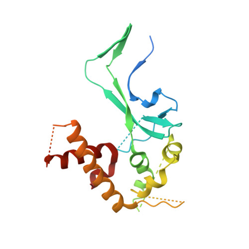
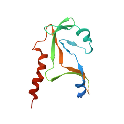
The Structure of the Human RNase H2 Complex Defines Key Interaction Interfaces Relevant to Enzyme Function and Human Disease.

Reijns, M.A.Bubeck, D.Gibson, L.C.Graham, S.C.Baillie, G.S.Jones, E.Y.Jackson, A.P.

(2011) J Biol Chem 286: 10530-10539

  • DOI: https://doi.org/10.1074/jbc.M110.177394
  • Primary Citation of Related Structures:  
    3P56, 3P5J

  • PubMed Abstract: 

    Ribonuclease H2 (RNase H2) is the major nuclear enzyme involved in the degradation of RNA/DNA hybrids and removal of ribonucleotides misincorporated in genomic DNA. Mutations in each of the three RNase H2 subunits have been implicated in a human auto-inflammatory disorder, Aicardi-Goutières Syndrome (AGS). To understand how mutations impact on RNase H2 function we determined the crystal structure of the human heterotrimer. In doing so, we correct several key regions of the previously reported murine RNase H2 atomic model and provide biochemical validation for our structural model. Our results provide new insights into how the subunits are arranged to form an enzymatically active complex. In particular, we establish that the RNASEH2A C terminus is a eukaryotic adaptation for binding the two accessory subunits, with residues within it required for enzymatic activity. This C-terminal extension interacts with the RNASEH2C C terminus and both are necessary to form a stable, enzymatically active heterotrimer. Disease mutations cluster at this interface between all three subunits, destabilizing the complex and/or impairing enzyme activity. Altogether, we locate 25 out of 29 residues mutated in AGS patients, establishing a firm basis for future investigations into disease pathogenesis and function of the RNase H2 enzyme.


  • Organizational Affiliation

    Medical Research Council Human Genetics Unit, Institute of Genetics and Molecular Medicine, Western General Hospital, Edinburgh EH4 2XU, United Kingdom.


Macromolecules
Find similar proteins by:  (by identity cutoff)  |  3D Structure
Entity ID: 1
MoleculeChains Sequence LengthOrganismDetailsImage
Ribonuclease H2 subunit A
A, D
299Homo sapiensMutation(s): 2 
Gene Names: RNASEH2ARNASEHIRNHIA
EC: 3.1.26.4
UniProt & NIH Common Fund Data Resources
Find proteins for O75792 (Homo sapiens)
Explore O75792 
Go to UniProtKB:  O75792
PHAROS:  O75792
GTEx:  ENSG00000104889 
Entity Groups  
Sequence Clusters30% Identity50% Identity70% Identity90% Identity95% Identity100% Identity
UniProt GroupO75792
Sequence Annotations
Expand
  • Reference Sequence
Find similar proteins by:  (by identity cutoff)  |  3D Structure
Entity ID: 2
MoleculeChains Sequence LengthOrganismDetailsImage
Ribonuclease H2 subunit B
B, E
237Homo sapiensMutation(s): 0 
Gene Names: DLEU8RNASEH2B
UniProt & NIH Common Fund Data Resources
Find proteins for Q5TBB1 (Homo sapiens)
Explore Q5TBB1 
Go to UniProtKB:  Q5TBB1
PHAROS:  Q5TBB1
GTEx:  ENSG00000136104 
Entity Groups  
Sequence Clusters30% Identity50% Identity70% Identity90% Identity95% Identity100% Identity
UniProt GroupQ5TBB1
Sequence Annotations
Expand
  • Reference Sequence
Find similar proteins by:  (by identity cutoff)  |  3D Structure
Entity ID: 3
MoleculeChains Sequence LengthOrganismDetailsImage
Ribonuclease H2 subunit C
C, F
164Homo sapiensMutation(s): 0 
Gene Names: AYP1RNASEH2C
UniProt & NIH Common Fund Data Resources
Find proteins for Q8TDP1 (Homo sapiens)
Explore Q8TDP1 
Go to UniProtKB:  Q8TDP1
PHAROS:  Q8TDP1
GTEx:  ENSG00000172922 
Entity Groups  
Sequence Clusters30% Identity50% Identity70% Identity90% Identity95% Identity100% Identity
UniProt GroupQ8TDP1
Sequence Annotations
Expand
  • Reference Sequence
Experimental Data & Validation

Experimental Data

  • Method: X-RAY DIFFRACTION
  • Resolution: 4.06 Å
  • R-Value Free: 0.379 
  • R-Value Work: 0.374 
  • R-Value Observed: 0.375 
  • Space Group: C 1 2 1
Unit Cell:
Length ( Å )Angle ( ˚ )
a = 212.238α = 90
b = 42.302β = 98.11
c = 186.95γ = 90
Software Package:
Software NamePurpose
DENZOdata reduction
SCALEPACKdata scaling
MOLREPphasing
BUSTER-TNTrefinement
PDB_EXTRACTdata extraction
ADSCdata collection
BUSTERrefinement

Structure Validation

View Full Validation Report


Entry History 

Deposition Data

Revision History  (Full details and data files)

  • Version 1.0: 2010-11-17
    Type: Initial release
  • Version 1.1: 2011-07-13
    Changes: Version format compliance
  • Version 1.2: 2023-09-06
    Changes: Data collection, Database references, Refinement description