3OXO

Succinyl-CoA:3-ketoacid CoA transferase from pig heart covalently bound to CoA


Experimental Data Snapshot

  • Method: X-RAY DIFFRACTION
  • Resolution: 2.30 Å
  • R-Value Free: 0.273 
  • R-Value Work: 0.245 
  • R-Value Observed: 0.247 

wwPDB Validation   3D Report Full Report


Ligand Structure Quality Assessment 


This is version 1.2 of the entry. See complete history


Literature

Catalytic role of the conformational change in succinyl-CoA:3-oxoacid CoA transferase on binding CoA.

Fraser, M.E.Hayakawa, K.Brown, W.D.

(2010) Biochemistry 49: 10319-10328

  • DOI: https://doi.org/10.1021/bi100659s
  • Primary Citation of Related Structures:  
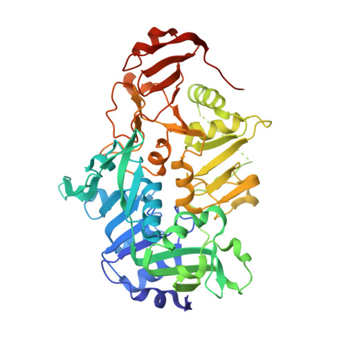
    3OXO

  • PubMed Abstract: 

    Catalysis by succinyl-CoA:3-oxoacid CoA transferase proceeds through a thioester intermediate in which CoA is covalently linked to the enzyme. To determine the conformation of the thioester intermediate, crystals of the pig enzyme were grown in the presence of the substrate acetoacetyl-CoA. X-ray diffraction data show the enzyme in both the free form and covalently bound to CoA via Glu305. In the complex, the protein adopts a conformation in which residues 267-275, 280-287, 357-373, and 398-477 have shifted toward Glu305, closing the enzyme around the thioester. Enzymes provide catalysis by stabilizing the transition state relative to complexes with substrates or products. In this case, the conformational change allows the enzyme to interact with parts of CoA distant from the reactive thiol while the thiol is covalently linked to the enzyme. The enzyme forms stabilizing interactions with both the nucleotide and pantoic acid portions of CoA, while the interactions with the amide groups of the pantetheine portion are poor. The results shed light on how the enzyme uses the binding energy for groups remote from the active center of CoA to destabilize atoms closer to the active center, leading to acceleration of the reaction by the enzyme.


  • Organizational Affiliation

    Department of Biological Sciences, University of Calgary, 2500 University Drive NW, Calgary, Alberta, Canada. frasm@ucalgary.ca


Macromolecules
Find similar proteins by:  (by identity cutoff)  |  3D Structure
Entity ID: 1
MoleculeChains Sequence LengthOrganismDetailsImage
Succinyl-CoA:3-ketoacid-coenzyme A transferase 1, mitochondrial
A, B, C, D, E
A, B, C, D, E, F, G, H
488Sus scrofaMutation(s): 0 
Gene Names: OXCTOXCT1SCOT
EC: 2.8.3.5
UniProt
Find proteins for Q29551 (Sus scrofa)
Explore Q29551 
Go to UniProtKB:  Q29551
Entity Groups  
Sequence Clusters30% Identity50% Identity70% Identity90% Identity95% Identity100% Identity
UniProt GroupQ29551
Sequence Annotations
Expand
  • Reference Sequence
Experimental Data & Validation

Experimental Data

  • Method: X-RAY DIFFRACTION
  • Resolution: 2.30 Å
  • R-Value Free: 0.273 
  • R-Value Work: 0.245 
  • R-Value Observed: 0.247 
  • Space Group: P 1
Unit Cell:
Length ( Å )Angle ( ˚ )
a = 69.121α = 89.6
b = 107.133β = 80.21
c = 134.565γ = 75.13
Software Package:
Software NamePurpose
ADSCdata collection
AMoREphasing
REFMACrefinement
MOSFLMdata reduction
SCALAdata scaling

Structure Validation

View Full Validation Report


Ligand Structure Quality Assessment 


Entry History 

Deposition Data

  • Released Date: 2010-11-10 
  • Deposition Author(s): Fraser, M.E.

Revision History  (Full details and data files)

  • Version 1.0: 2010-11-10
    Type: Initial release
  • Version 1.1: 2011-07-13
    Changes: Version format compliance
  • Version 1.2: 2023-09-06
    Changes: Data collection, Database references, Derived calculations, Refinement description