Contribution of the 80s loop of HIV-1 protease to the multidrug-resistance mechanism: crystallographic study of MDR769 HIV-1 protease variants.
Yedidi, R.S., Proteasa, G., Martinez, J.L., Vickrey, J.F., Martin, P.D., Wawrzak, Z., Liu, Z., Kovari, I.A., Kovari, L.C.(2011) Acta Crystallogr D Biol Crystallogr 67: 524-532
- PubMed: 21636892
- DOI: https://doi.org/10.1107/S0907444911011541
- Primary Citation of Related Structures:
3OQ7, 3OQA, 3OQD, 3PJ6 - PubMed Abstract:
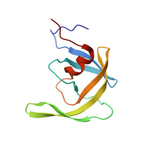
The flexible flaps and the 80s loops (Pro79-Ile84) of HIV-1 protease are crucial in inhibitor binding. Previously, it was reported that the crystal structure of multidrug-resistant 769 (MDR769) HIV-1 protease shows a wide-open conformation of the flaps owing to conformational rigidity acquired by the accumulation of mutations. In the current study, the effect of mutations on the conformation of the 80s loop of MDR769 HIV-1 protease variants is reported. Alternate conformations of Pro81 (proline switch) with a root-mean-square deviation of 3-4.8 Å in the C(α) atoms of the I10V mutant and a side chain with a `flipped-out' conformation in the A82F mutant cause distortion in the S1/S1' binding pockets that affects inhibitor binding. The A82S and A82T mutants show local changes in the electrostatics of inhibitor binding owing to the mutation from nonpolar to polar residues. In summary, the crystallographic studies of four variants of MDR769 HIV-1 protease presented in this article provide new insights towards understanding the drug-resistance mechanism as well as a basis for design of future protease inhibitors with enhanced potency.
Organizational Affiliation:
Department of Biochemistry and Molecular Biology, School of Medicine, Wayne State University, 540 East Canfield Avenue, Detroit, MI 48201, USA.