3OEO

The crystal structure E. coli Spy


Experimental Data Snapshot

  • Method: X-RAY DIFFRACTION
  • Resolution: 2.70 Å
  • R-Value Free: 0.300 
  • R-Value Work: 0.252 
  • R-Value Observed: 0.255 

wwPDB Validation   3D Report Full Report


This is version 1.2 of the entry. See complete history


Literature

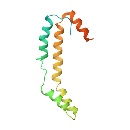
The crystal structure Escherichia coli Spy.

Kwon, E.Kim, D.Y.Gross, C.A.Gross, J.D.Kim, K.K.

(2010) Protein Sci 19: 2252-2259

  • DOI: https://doi.org/10.1002/pro.489
  • Primary Citation of Related Structures:  
    3OEO

  • PubMed Abstract: 

    Escherichia coli spheroplast protein y (EcSpy) is a small periplasmic protein that is homologous with CpxP, an inhibitor of the extracytoplasmic stress response. Stress conditions such as spheroplast formation induce the expression of Spy via the Cpx or the Bae two-component systems in E. coli, though the function of Spy is unknown. Here, we report the crystal structure of EcSpy, which reveals a long kinked hairpin-like structure of four α-helices that form an antiparallel dimer. The dimer contains a curved oval shape with a highly positively charged concave surface that may function as a ligand binding site. Sequence analysis reveals that Spy is highly conserved over the Enterobacteriaceae family. Notably, three conserved regions that contain identical residues and two LTxxQ motifs are placed at the horizontal end of the dimer structure, stabilizing the overall fold. CpxP also contains the conserved sequence motifs and has a predicted secondary structure similar to Spy, suggesting that Spy and CpxP likely share the same fold.


  • Organizational Affiliation

    Department of Molecular Cell Biology, Samsung Biomedical Research Institute, Sungkyunkwan University School of Medicine, Suwon, Korea.


Macromolecules
Find similar proteins by:  (by identity cutoff)  |  3D Structure
Entity ID: 1
MoleculeChains Sequence LengthOrganismDetailsImage
Spheroplast protein Y
A, B, C, D
138Escherichia coliMutation(s): 0 
Gene Names: spyb1743JW1732
UniProt
Find proteins for C6ECL5 (Escherichia coli (strain B / BL21-DE3))
Explore C6ECL5 
Go to UniProtKB:  C6ECL5
Entity Groups  
Sequence Clusters30% Identity50% Identity70% Identity90% Identity95% Identity100% Identity
UniProt GroupC6ECL5
Sequence Annotations
Expand
  • Reference Sequence
Small Molecules
Ligands 1 Unique
IDChains Name / Formula / InChI Key2D Diagram3D Interactions
CD
Query on CD

Download Ideal Coordinates CCD File 
E [auth A]
F [auth A]
G [auth A]
H [auth A]
I [auth A]
E [auth A],
F [auth A],
G [auth A],
H [auth A],
I [auth A],
J [auth A],
K [auth B],
L [auth B],
M [auth B],
N [auth B],
O [auth B],
P [auth C],
Q [auth C],
R [auth C],
S [auth C],
T [auth C],
U [auth C],
V [auth C],
W [auth D],
X [auth D],
Y [auth D],
Z [auth D]
CADMIUM ION
Cd
WLZRMCYVCSSEQC-UHFFFAOYSA-N
Experimental Data & Validation

Experimental Data

  • Method: X-RAY DIFFRACTION
  • Resolution: 2.70 Å
  • R-Value Free: 0.300 
  • R-Value Work: 0.252 
  • R-Value Observed: 0.255 
  • Space Group: P 32
Unit Cell:
Length ( Å )Angle ( ˚ )
a = 69.195α = 90
b = 69.195β = 90
c = 126.347γ = 120
Software Package:
Software NamePurpose
SCALEPACKdata scaling
REFMACrefinement
PDB_EXTRACTdata extraction
ADSCdata collection
HKL-2000data reduction
SOLVEphasing

Structure Validation

View Full Validation Report


Entry History 

Deposition Data

Revision History  (Full details and data files)

  • Version 1.0: 2010-09-22
    Type: Initial release
  • Version 1.1: 2011-07-13
    Changes: Version format compliance
  • Version 1.2: 2024-02-21
    Changes: Data collection, Database references, Derived calculations