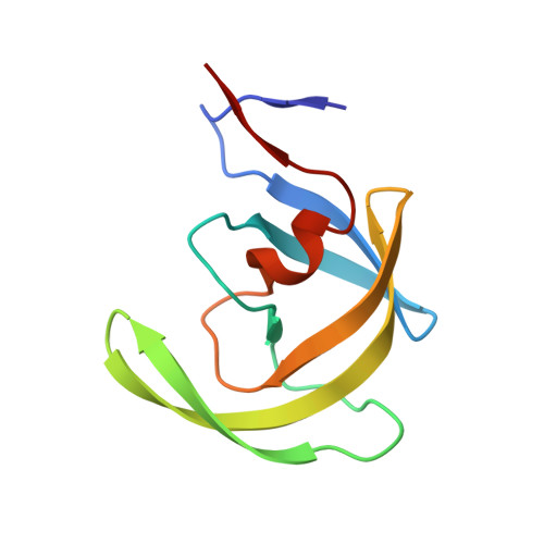
3O99

Crystal Structure of wild-type HIV-1 Protease in complex with kd13


Experimental Data Snapshot

  • Method: X-RAY DIFFRACTION
  • Resolution: 1.95 Å
  • R-Value Free: 0.224 
  • R-Value Work: 0.177 
  • R-Value Observed: 0.179 

wwPDB Validation   3D Report Full Report


Ligand Structure Quality Assessment 


This is version 1.2 of the entry. See complete history


Literature

Substrate envelope-designed potent HIV-1 protease inhibitors to avoid drug resistance.

Nalam, M.N.Ali, A.Reddy, G.S.Cao, H.Anjum, S.G.Altman, M.D.Yilmaz, N.K.Tidor, B.Rana, T.M.Schiffer, C.A.

(2013) Chem Biol 20: 1116-1124

  • DOI: https://doi.org/10.1016/j.chembiol.2013.07.014
  • Primary Citation of Related Structures:  
    3O99, 3O9A, 3O9B, 3O9C, 3O9D, 3O9E, 3O9F, 3O9G, 3O9H, 3O9I

  • PubMed Abstract: 

    The rapid evolution of HIV under selective drug pressure has led to multidrug resistant (MDR) strains that evade standard therapies. We designed highly potent HIV-1 protease inhibitors (PIs) using the substrate envelope model, which confines inhibitors within the consensus volume of natural substrates, providing inhibitors less susceptible to resistance because a mutation affecting such inhibitors will simultaneously affect viral substrate processing. The designed PIs share a common chemical scaffold but utilize various moieties that optimally fill the substrate envelope, as confirmed by crystal structures. The designed PIs retain robust binding to MDR protease variants and display exceptional antiviral potencies against different clades of HIV as well as a panel of 12 drug-resistant viral strains. The substrate envelope model proves to be a powerful strategy to develop potent and robust inhibitors that avoid drug resistance.


  • Organizational Affiliation

    Department of Biochemistry and Molecular Pharmacology, University of Massachusetts Medical School, Worcester, MA 01605, USA.


Macromolecules
Find similar proteins by:  (by identity cutoff)  |  3D Structure
Entity ID: 1
MoleculeChains Sequence LengthOrganismDetailsImage
Pol polyprotein
A, B
99Human immunodeficiency virus 1Mutation(s): 1 
Gene Names: gag-polpol
UniProt
Find proteins for Q90K99 (Human immunodeficiency virus 1)
Explore Q90K99 
Go to UniProtKB:  Q90K99
Entity Groups  
Sequence Clusters30% Identity50% Identity70% Identity90% Identity95% Identity100% Identity
UniProt GroupQ90K99
Sequence Annotations
Expand
  • Reference Sequence
Small Molecules
Ligands 3 Unique
IDChains Name / Formula / InChI Key2D Diagram3D Interactions
K13
Query on K13

Download Ideal Coordinates CCD File 
D [auth A](3R,3aS,6aR)-hexahydrofuro[2,3-b]furan-3-yl [(1S,2R)-3-{[(4-aminophenyl)sulfonyl][(2S)-2-methylbutyl]amino}-1-benzyl-2-hydroxypropyl]carbamate
C28 H39 N3 O7 S
NKLRQHQHYBFPSD-DRMLODCJSA-N
PO4
Query on PO4

Download Ideal Coordinates CCD File 
C [auth A],
E [auth B]
PHOSPHATE ION
O4 P
NBIIXXVUZAFLBC-UHFFFAOYSA-K
ACT
Query on ACT

Download Ideal Coordinates CCD File 
F [auth B]ACETATE ION
C2 H3 O2
QTBSBXVTEAMEQO-UHFFFAOYSA-M
Binding Affinity Annotations 
IDSourceBinding Affinity
K13 BindingDB:  3O99 Ki: min: 0.01, max: 0.01 (nM) from 2 assay(s)
PDBBind:  3O99 Ki: 0.02 (nM) from 1 assay(s)
Experimental Data & Validation

Experimental Data

  • Method: X-RAY DIFFRACTION
  • Resolution: 1.95 Å
  • R-Value Free: 0.224 
  • R-Value Work: 0.177 
  • R-Value Observed: 0.179 
  • Space Group: P 21 21 21
Unit Cell:
Length ( Å )Angle ( ˚ )
a = 50.774α = 90
b = 57.885β = 90
c = 61.752γ = 90
Software Package:
Software NamePurpose
DENZOdata reduction
SCALEPACKdata scaling
REFMACrefinement
PDB_EXTRACTdata extraction
AMoREphasing

Structure Validation

View Full Validation Report


Ligand Structure Quality Assessment 


Entry History 

Deposition Data

Revision History  (Full details and data files)

  • Version 1.0: 2011-08-10
    Type: Initial release
  • Version 1.1: 2013-11-27
    Changes: Database references
  • Version 1.2: 2023-09-06
    Changes: Data collection, Database references, Derived calculations, Refinement description