Biochemical and structural characterization of bisubstrate inhibitors of BasE, the self-standing nonribosomal peptide synthetase adenylate-forming enzyme of acinetobactin synthesis.
Drake, E.J., Duckworth, B.P., Neres, J., Aldrich, C.C., Gulick, A.M.(2010) Biochemistry 49: 9292-9305
- PubMed: 20853905
- DOI: https://doi.org/10.1021/bi101226n
- Primary Citation of Related Structures:
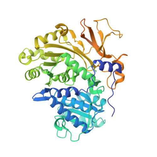
3O82, 3O83, 3O84 - PubMed Abstract:
The human pathogen Acinetobacter baumannii produces a siderophore called acinetobactin that is derived from one molecule each of threonine, histidine, and 2,3-dihydroxybenzoic acid (DHB). The activity of several nonribosomal peptide synthetase (NRPS) enzymes is used to combine the building blocks into the final molecule. The acinetobactin synthesis pathway initiates with a self-standing adenylation enzyme, BasE, that activates the DHB molecule and covalently transfers it to the pantetheine cofactor of an aryl-carrier protein of BasF, a strategy that is shared with many siderophore-producing NRPS clusters. In this reaction, DHB reacts with ATP to form the aryl adenylate and pyrophosphate. In a second partial reaction, the DHB is transferred to the carrier protein. Inhibitors of BasE and related enzymes have been identified that prevent growth of bacteria on iron-limiting media. Recently, a new inhibitor of BasE has been identified via high-throughput screening using a fluorescence polarization displacement assay. We present here biochemical and structural studies to examine the binding mode of this inhibitor. The kinetics of the wild-type BasE enzyme is shown, and inhibition studies demonstrate that the new compound exhibits competitive inhibition against both ATP and 2,3-dihydroxybenzoate. Structural examination of BasE bound to this inhibitor illustrates a novel binding mode in which the phenyl moiety partially fills the enzyme pantetheine binding tunnel. Structures of rationally designed bisubstrate inhibitors are also presented.
Organizational Affiliation:
Hauptman-Woodward Institute and Department of Structural Biology, University at Buffalo, Buffalo, NY 14203-1102, USA.