3O4F

Crystal Structure of Spermidine Synthase from E. coli


Experimental Data Snapshot

  • Method: X-RAY DIFFRACTION
  • Resolution: 2.90 Å
  • R-Value Free: 0.241 
  • R-Value Work: 0.208 
  • R-Value Observed: 0.210 

wwPDB Validation   3D Report Full Report


This is version 1.3 of the entry. See complete history


Literature

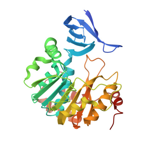
The crystal structure of Escherichia coli spermidine synthase SpeE reveals a unique substrate-binding pocket

Zhou, X.Chua, T.K.Tkaczuk, K.L.Bujnicki, J.M.Sivaraman, J.

(2010) J Struct Biol 169: 277-285

  • DOI: https://doi.org/10.1016/j.jsb.2009.12.024
  • Primary Citation of Related Structures:  
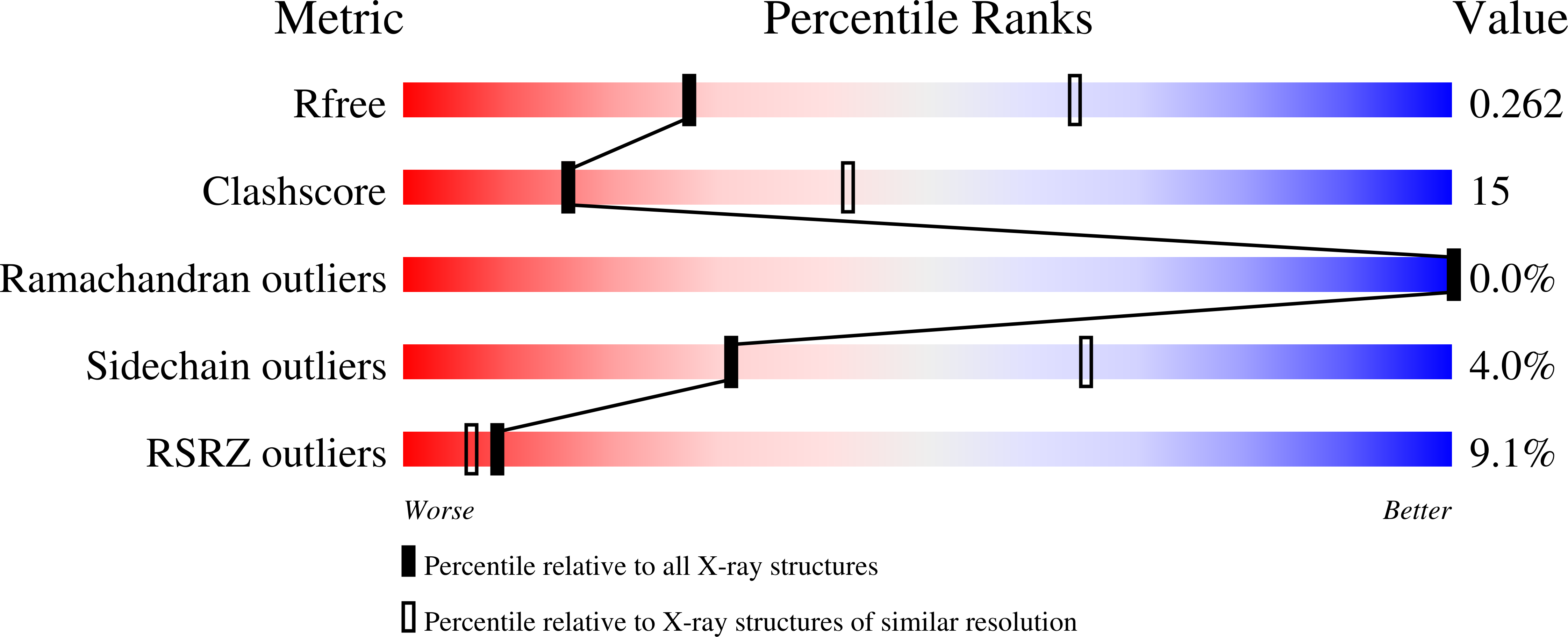
    3O4F

  • PubMed Abstract: 

    Polyamines are essential in all branches of life. Biosynthesis of spermidine, one of the most ubiquitous polyamines, is catalyzed by spermidine synthase (SpeE). Although the function of this enzyme from Escherichia coli has been thoroughly characterised, its structural details remain unknown. Here, we report the crystal structure of E. coli SpeE and study its interaction with the ligands by isothermal titration calorimetry and computational modelling. SpeE consists of two domains - a small N-terminal beta-strand domain, and a C-terminal catalytic domain that adopts a canonical methyltransferase (MTase) Rossmann fold. The protein forms a dimer in the crystal and in solution. Structural comparison of E. coli SpeE to its homologs reveals that it has a large and unique substrate-binding cleft that may account for its lower amine substrate specificity.


  • Organizational Affiliation

    Department of Biological Sciences, 14 Science Drive 4, National University of Singapore, Singapore 117543, Republic of Singapore.


Macromolecules
Find similar proteins by:  (by identity cutoff)  |  3D Structure
Entity ID: 1
MoleculeChains Sequence LengthOrganismDetailsImage
Spermidine synthase
A, B, C, D, E
A, B, C, D, E, F, G, H
294Escherichia coli K-12Mutation(s): 0 
Gene Names: SPEE
EC: 2.5.1.16
UniProt
Find proteins for P09158 (Escherichia coli (strain K12))
Explore P09158 
Go to UniProtKB:  P09158
Entity Groups  
Sequence Clusters30% Identity50% Identity70% Identity90% Identity95% Identity100% Identity
UniProt GroupP09158
Sequence Annotations
Expand
  • Reference Sequence
Small Molecules
Ligands 1 Unique
IDChains Name / Formula / InChI Key2D Diagram3D Interactions
SO4
Query on SO4

Download Ideal Coordinates CCD File 
AA [auth E]
BA [auth E]
CA [auth F]
DA [auth F]
EA [auth F]
AA [auth E],
BA [auth E],
CA [auth F],
DA [auth F],
EA [auth F],
FA [auth F],
GA [auth G],
HA [auth G],
I [auth A],
IA [auth G],
J [auth A],
JA [auth G],
K [auth A],
KA [auth G],
L [auth A],
LA [auth H],
M [auth B],
MA [auth H],
N [auth B],
O [auth C],
P [auth C],
Q [auth C],
R [auth C],
S [auth C],
T [auth C],
U [auth D],
V [auth D],
W [auth D],
X [auth D],
Y [auth E],
Z [auth E]
SULFATE ION
O4 S
QAOWNCQODCNURD-UHFFFAOYSA-L
Experimental Data & Validation

Experimental Data

  • Method: X-RAY DIFFRACTION
  • Resolution: 2.90 Å
  • R-Value Free: 0.241 
  • R-Value Work: 0.208 
  • R-Value Observed: 0.210 
  • Space Group: P 43
Unit Cell:
Length ( Å )Angle ( ˚ )
a = 123.11α = 90
b = 123.11β = 90
c = 210.01γ = 90
Software Package:
Software NamePurpose
REFMACrefinement

Structure Validation

View Full Validation Report


Entry History 

Deposition Data

Revision History  (Full details and data files)

  • Version 1.0: 2010-08-18
    Type: Initial release
  • Version 1.1: 2011-07-13
    Changes: Version format compliance
  • Version 1.2: 2022-04-13
    Changes: Database references, Derived calculations, Structure summary
  • Version 1.3: 2023-11-01
    Changes: Data collection, Refinement description