3O11

Anti-beta-amyloid antibody c706 fab in space group c2


Experimental Data Snapshot

  • Method: X-RAY DIFFRACTION
  • Resolution: 2.80 Å
  • R-Value Free: 0.304 
  • R-Value Work: 0.220 
  • R-Value Observed: 0.223 

wwPDB Validation   3D Report Full Report


This is version 2.2 of the entry. See complete history


Literature

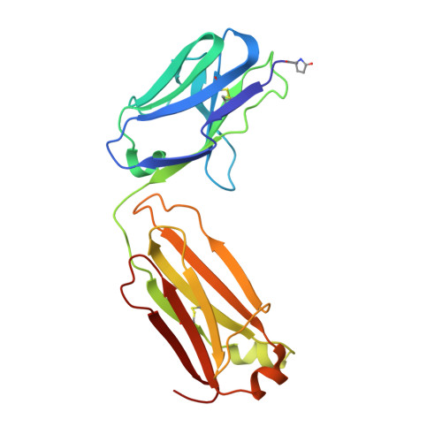
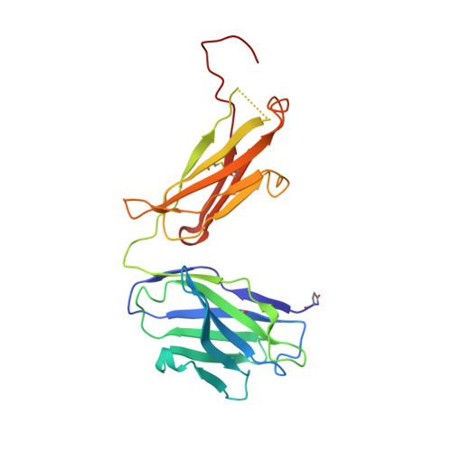
His-tag binding by antibody C706 mimics beta-amyloid recognition.

Teplyakov, A.Obmolova, G.Canziani, G.Zhao, Y.Gutshall, L.Jung, S.S.Gilliland, G.L.

(2011) J Mol Recognit 24: 570-575

  • DOI: https://doi.org/10.1002/jmr.1069
  • Primary Citation of Related Structures:  
    3MCL, 3O11

  • PubMed Abstract: 

    Alzheimer's disease is a progressive neurodegenerative disease characterized by extracellular deposits of β-amyloid (Aβ) plaques. Aggregation of the Aβ(42) peptide leading to plaque formation is believed to play a central role in Alzheimer's disease pathogenesis. Anti-Aβ monoclonal antibodies can reduce amyloid plaques and could possibly be used for immunotherapy. We have developed a monoclonal antibody C706, which recognizes the human Aβ peptide. Here we report the crystal structure of the antibody Fab fragment at 1.7 Å resolution. The structure was determined in two crystal forms, P2(1) and C2. Although the Fab was crystallized in the presence of Aβ(16), no peptide was observed in the crystals. The antigen-binding site is blocked by the hexahistidine tag of another Fab molecule in both crystal forms. The poly-His peptide in an extended conformation occupies a crevice between the light and heavy chains of the variable domain. Two consecutive histidines (His4-His5) stack against tryptophan residues in the central pocket of the antigen-binding surface. In addition, they form hydrogen bonds to the acidic residues at the bottom of the pocket. The mode of his-tag binding by C706 resembles the Aβ recognition by antibodies PFA1 and WO2. All three antibodies recognize the same immunodominant B-cell epitope of Aβ. By similarity, residues Phe-Arg-His of Aβ would be a major portion of the C706 epitope.


  • Organizational Affiliation

    Centocor R&D, 145 King of Prussia Road, Radnor, PA 19087, USA. ateplyak@its.jnj.com


Macromolecules
Find similar proteins by:  (by identity cutoff)  |  3D Structure
Entity ID: 1
MoleculeChains Sequence LengthOrganismDetailsImage
C706 LIGHT CHAIN variable region, Ig kappa chain C region chimeraA [auth L],
C [auth A]
212Mus musculusHomo sapiens
This entity is chimeric
Mutation(s): 0 
Gene Names: IGKC
UniProt & NIH Common Fund Data Resources
Find proteins for P01834 (Homo sapiens)
Explore P01834 
Go to UniProtKB:  P01834
PHAROS:  P01834
Entity Groups  
Sequence Clusters30% Identity50% Identity70% Identity90% Identity95% Identity100% Identity
UniProt GroupP01834
Sequence Annotations
Expand
  • Reference Sequence
Find similar proteins by:  (by identity cutoff)  |  3D Structure
Entity ID: 2
MoleculeChains Sequence LengthOrganismDetailsImage
C706 HEAVY CHAIN variable region, Ig gamma-1 chain C region chimeraB [auth H],
D [auth B]
228Mus musculusHomo sapiens
This entity is chimeric
Mutation(s): 0 
Gene Names: IGHG1
UniProt & NIH Common Fund Data Resources
Find proteins for P01857 (Homo sapiens)
Explore P01857 
Go to UniProtKB:  P01857
PHAROS:  P01857
GTEx:  ENSG00000211896 
Entity Groups  
Sequence Clusters30% Identity50% Identity70% Identity90% Identity95% Identity100% Identity
UniProt GroupP01857
Sequence Annotations
Expand
  • Reference Sequence
Small Molecules
Modified Residues  1 Unique
IDChains TypeFormula2D DiagramParent
PCA
Query on PCA
A [auth L],
C [auth A]
L-PEPTIDE LINKINGC5 H7 N O3GLN
Experimental Data & Validation

Experimental Data

  • Method: X-RAY DIFFRACTION
  • Resolution: 2.80 Å
  • R-Value Free: 0.304 
  • R-Value Work: 0.220 
  • R-Value Observed: 0.223 
  • Space Group: C 1 2 1
Unit Cell:
Length ( Å )Angle ( ˚ )
a = 136.04α = 90
b = 45.02β = 92.69
c = 158.2γ = 90
Software Package:
Software NamePurpose
CrystalCleardata collection
AMoREphasing
REFMACrefinement
d*TREKdata reduction
d*TREKdata scaling

Structure Validation

View Full Validation Report


Entry History 

Deposition Data

Revision History  (Full details and data files)

  • Version 1.0: 2010-09-08
    Type: Initial release
  • Version 1.1: 2011-07-13
    Changes: Version format compliance
  • Version 1.2: 2012-09-05
    Changes: Derived calculations
  • Version 1.3: 2017-08-16
    Changes: Refinement description, Source and taxonomy
  • Version 2.0: 2019-12-25
    Changes: Database references, Derived calculations, Polymer sequence
  • Version 2.1: 2023-07-26
    Changes: Database references
  • Version 2.2: 2023-09-06
    Changes: Data collection, Refinement description