


Crystal structure of glutathione reductase from Bartonella henselae
Seattle Structural Genomics Center for Infectious Disease (SSGCID), Abendroth, J., Edwards, T.E., Staker, B.To be published.
Experimental Data Snapshot
Entity ID: 1 | |||||
|---|---|---|---|---|---|
| Molecule | Chains | Sequence Length | Organism | Details | Image |
| Glutathione reductase | 484 | Bartonella henselae str. Houston-1 | Mutation(s): 0 Gene Names: gor, BH06430 EC: 1.8.1.7 | 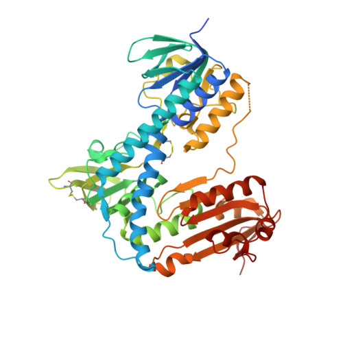 | |
UniProt | |||||
Find proteins for A0A0H3LWY9 (Bartonella henselae (strain ATCC 49882 / DSM 28221 / CCUG 30454 / Houston 1)) Explore A0A0H3LWY9 Go to UniProtKB: A0A0H3LWY9 | |||||
Entity Groups | |||||
| Sequence Clusters | 30% Identity50% Identity70% Identity90% Identity95% Identity100% Identity | ||||
| UniProt Group | A0A0H3LWY9 | ||||
Sequence AnnotationsExpand | |||||
| |||||
| Ligands 2 Unique | |||||
|---|---|---|---|---|---|
| ID | Chains | Name / Formula / InChI Key | 2D Diagram | 3D Interactions | |
| FAD Query on FAD | E [auth A], H [auth B] | FLAVIN-ADENINE DINUCLEOTIDE C27 H33 N9 O15 P2 VWWQXMAJTJZDQX-UYBVJOGSSA-N |  | ||
| CL Query on CL | C [auth A], D [auth A], F [auth B], G [auth B] | CHLORIDE ION Cl VEXZGXHMUGYJMC-UHFFFAOYSA-M |  | ||
| Length ( Å ) | Angle ( ˚ ) |
|---|---|
| a = 84.47 | α = 90 |
| b = 64.65 | β = 107.24 |
| c = 90.19 | γ = 90 |
| Software Name | Purpose |
|---|---|
| StructureStudio | data collection |
| PHASER | phasing |
| REFMAC | refinement |
| XDS | data reduction |
| XSCALE | data scaling |
RCSB PDB (citation) is hosted by
RCSB PDB is a member of the

