3NF3

Crystal structure of BoNT/A LC with JTH-NB-7239 peptide


Experimental Data Snapshot

  • Method: X-RAY DIFFRACTION
  • Resolution: 2.40 Å
  • R-Value Free: 0.231 
  • R-Value Work: 0.183 
  • R-Value Observed: 0.185 

wwPDB Validation   3D Report Full Report


This is version 1.4 of the entry. See complete history


Literature

Iterative structure-based peptide-like inhibitor design against the botulinum neurotoxin serotype A.

Zuniga, J.E.Hammill, J.T.Drory, O.Nuss, J.E.Burnett, J.C.Gussio, R.Wipf, P.Bavari, S.Brunger, A.T.

(2010) PLoS One 5: e11378-e11378

  • DOI: https://doi.org/10.1371/journal.pone.0011378
  • Primary Citation of Related Structures:  
    3NF3

  • PubMed Abstract: 

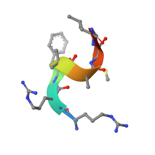
    The botulinum neurotoxin serotype A light chain (BoNT/A LC) protease is the catalytic component responsible for the neuroparalysis that is characteristic of the disease state botulism. Three related peptide-like molecules (PLMs) were designed using previous information from co-crystal structures, synthesized, and assayed for in vitro inhibition against BoNT/A LC. Our results indicate these PLMS are competitive inhibitors of the BoNT/A LC protease and their K(i) values are in the nM-range. A co-crystal structure for one of these inhibitors was determined and reveals that the PLM, in accord with the goals of our design strategy, simultaneously involves both ionic interactions via its P1 residue and hydrophobic contacts by means of an aromatic group in the P2' position. The PLM adopts a helical conformation similar to previously determined co-crystal structures of PLMs, although there are also major differences to these other structures such as contacts with specific BoNT/A LC residues. Our structure further demonstrates the remarkable plasticity of the substrate binding cleft of the BoNT/A LC protease and provides a paradigm for iterative structure-based design and development of BoNT/A LC inhibitors.


  • Organizational Affiliation

    Howard Hughes Medical Institute and Department of Molecular and Cellular Physiology, Stanford University, Stanford, California, United States of America.


Macromolecules
Find similar proteins by:  (by identity cutoff)  |  3D Structure
Entity ID: 1
MoleculeChains Sequence LengthOrganismDetailsImage
BoNT/A425Clostridium botulinumMutation(s): 0 
Gene Names: bont/aboNT/Abonta
UniProt
Find proteins for Q7B8V4 (Clostridium botulinum)
Explore Q7B8V4 
Go to UniProtKB:  Q7B8V4
Entity Groups  
Sequence Clusters30% Identity50% Identity70% Identity90% Identity95% Identity100% Identity
UniProt GroupQ7B8V4
Sequence Annotations
Expand
  • Reference Sequence

Find similar proteins by:  Sequence   |   3D Structure  

Entity ID: 2
MoleculeChains Sequence LengthOrganismDetailsImage
JTH-NB72-39 inhibitorB [auth C]8N/AMutation(s): 0 
Sequence Annotations
Expand
  • Reference Sequence
Small Molecules
Modified Residues  1 Unique
IDChains TypeFormula2D DiagramParent
AIB
Query on AIB
B [auth C]L-PEPTIDE LINKINGC4 H9 N O2ALA
Biologically Interesting Molecules (External Reference) 1 Unique
Experimental Data & Validation

Experimental Data

  • Method: X-RAY DIFFRACTION
  • Resolution: 2.40 Å
  • R-Value Free: 0.231 
  • R-Value Work: 0.183 
  • R-Value Observed: 0.185 
  • Space Group: P 21 21 2
Unit Cell:
Length ( Å )Angle ( ˚ )
a = 56.1α = 90
b = 189.6β = 90
c = 41.51γ = 90
Software Package:
Software NamePurpose
Blu-Icedata collection
PHASERphasing
PHENIXrefinement
HKL-2000data reduction
HKL-2000data scaling

Structure Validation

View Full Validation Report


Entry History 

Deposition Data

  • Released Date: 2010-07-21 
  • Deposition Author(s): Zuniga, J.E.

Revision History  (Full details and data files)

  • Version 1.0: 2010-07-21
    Type: Initial release
  • Version 1.1: 2011-07-13
    Changes: Version format compliance
  • Version 1.2: 2012-12-12
    Changes: Other
  • Version 1.3: 2023-09-06
    Changes: Data collection, Database references, Derived calculations, Refinement description
  • Version 1.4: 2023-11-22
    Changes: Data collection