Gradual adaptive changes of a protein facing high salt concentrations.
Coquelle, N., Talon, R., Juers, D.H., Girard, E., Kahn, R., Madern, D.(2010) J Mol Biol 404: 493-505
- PubMed: 20888835
- DOI: https://doi.org/10.1016/j.jmb.2010.09.055
- Primary Citation of Related Structures:
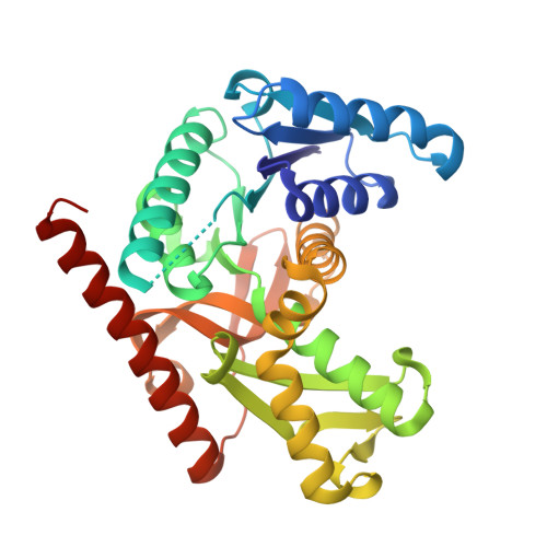
3NEP - PubMed Abstract:
Several experimental techniques were applied to unravel fine molecular details of protein adaptation to high salinity. We compared four homologous enzymes, which suggested a new halo-adaptive state in the process of molecular adaptation to high-salt conditions. Together with comparative functional studies, the structure of malate dehydrogenase from the eubacterium Salinibacter ruber shows that the enzyme shares characteristics of a halo-adapted archaea-bacterial enzyme and of non-halo-adapted enzymes from other eubacterial species. The S. ruber enzyme is active at the high physiological concentrations of KCl but, unlike typical halo-adapted enzymes, remains folded and active at low salt concentrations. Structural aspects of the protein, including acidic residues at the surface, solvent-exposed hydrophobic surface, and buried hydrophobic surface, place it between the typical halo-adapted and non-halo-adapted proteins. The enzyme lacks inter-subunit ion-binding sites often seen in halo-adapted enzymes. These observations permit us to suggest an evolutionary pathway that is highlighted by subtle trade-offs to achieve an optimal compromise among solubility, stability, and catalytic activity.
Organizational Affiliation:
IBS, Institut de Biologie Structurale Jean-Pierre Ébel, Extremophilic and Large Molecular Assemblies Team, UMR 5075, CEA, CNRS, Université Joseph Fourier, 41 rue Jules Horowitz, F-38027 Grenoble, France.