Conformational exchange is critical for the productivity of an oxidative folding intermediate with buried free cysteines.
Gross, G., Gallopin, M., Vandame, M., Couprie, J., Stura, E., Zinn-Justin, S., Drevet, P.(2010) J Mol Biol 403: 299-312
- PubMed: 20804768
- DOI: https://doi.org/10.1016/j.jmb.2010.07.048
- Primary Citation of Related Structures:
3NDS - PubMed Abstract:
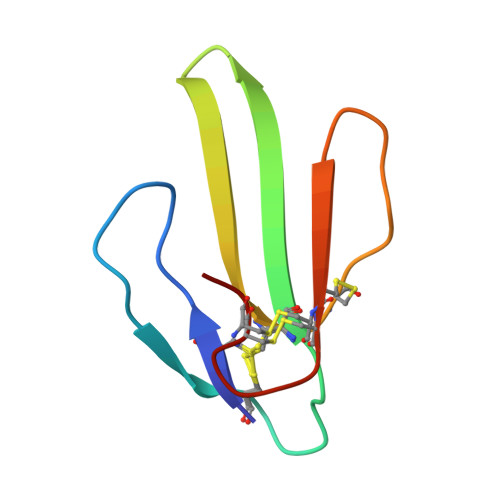
Much has been learned about the folding of proteins from comparative studies of the folding of proteins that are related in sequence and structure. Observation of the effects of mutations helps account for sequence-specific properties and large variations in folding rates observed in homologous proteins, which are not explained by structure-derived descriptions. The folding kinetics of variants of a β-stranded protein, toxin α from Naja nigricollis, depends on the length of their loop lk1. These proteins, named Tox60, Tox61, and Tox62, contain four disulfide bonds. We show that their oxidative refolding pathways are similar. Differences in these pathways are restricted to the last step of the reaction, that is, the closure of the last disulfide. At this step, two species of three-disulfide intermediates are observed: intermediate C lacking the B3 disulfide and intermediate D lacking the B2 disulfide. Surprisingly, D is the most productive intermediate for Tox61 despite the low accessibility of its free cysteines. However, in the case of Tox62, its conversion efficiency drops by 2 orders of magnitude and C becomes the most productive intermediate. NMR was used in order to study the structural dynamics of each of these intermediates. Both three-disulfide intermediates of Tox61 exist in two forms, exchanging on the 1- to 100-ms scale. One of these forms is structurally very close to the native Tox61, whereas the other is always significantly more flexible on a picosecond-to-nanosecond timescale. On the other hand, in the case of Tox62, the three-disulfide intermediates only show a native-like structure. The higher conformational heterogeneity of Tox61 intermediate D allows an increased accessibility of its free cysteines to oxidative agents, which explains its faster native disulfide formation. Thus, residue deletion in loop lk1 probably abrogates stabilizing intramolecular interactions, creates conformational heterogeneity, and increases the folding rate of Tox60 and Tox61 compared to Tox62.
Organizational Affiliation:
CEA/DSV/iBiTEC-S/SBIGeM, F-91191 Gif sur Yvette Cedex, France.