


X-ray Crystal Structures of TREX1 3' Exonuclease Autoimmune Disease Mutants
Bailey, S.L., Harvey, S., Perrino, F.W., Hollis, T.To be published.
Experimental Data Snapshot
wwPDB Validation 3D Report Full Report

Entity ID: 1 | |||||
|---|---|---|---|---|---|
| Molecule | Chains | Sequence Length | Organism | Details | Image |
| Three prime repair exonuclease 1 | A [auth B], B [auth A] | 242 | Mus musculus | Mutation(s): 0 Gene Names: Trex1 EC: 3.1.11.2 | 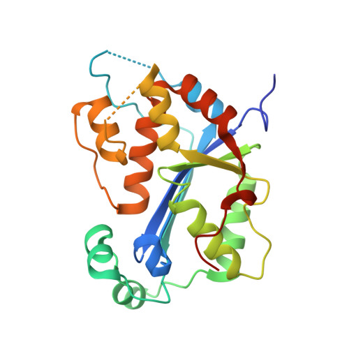 |
UniProt | |||||
Find proteins for Q91XB0 (Mus musculus) Explore Q91XB0 Go to UniProtKB: Q91XB0 | |||||
Entity Groups | |||||
| Sequence Clusters | 30% Identity50% Identity70% Identity90% Identity95% Identity100% Identity | ||||
| UniProt Group | Q91XB0 | ||||
Sequence AnnotationsExpand | |||||
| |||||
| Length ( Å ) | Angle ( ˚ ) |
|---|---|
| a = 64.16 | α = 90 |
| b = 85.72 | β = 90 |
| c = 100.27 | γ = 90 |
| Software Name | Purpose |
|---|---|
| CrystalClear | data collection |
| PHASER | phasing |
| PHENIX | refinement |
| d*TREK | data reduction |
| d*TREK | data scaling |
RCSB PDB (citation) is hosted by
RCSB PDB is a member of the

