Plasticity of Cytochrome P450 2B4 as Investigated by Hydrogen-Deuterium Exchange Mass Spectrometry and X-ray Crystallography.
Wilderman, P.R., Shah, M.B., Liu, T., Li, S., Hsu, S., Roberts, A.G., Goodlett, D.R., Zhang, Q., Woods, V.L., Stout, C.D., Halpert, J.R.(2010) J Biol Chem 285: 38602-38611
- PubMed: 20880847
- DOI: https://doi.org/10.1074/jbc.M110.180646
- Primary Citation of Related Structures:
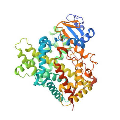
3MVR - PubMed Abstract:
Crystal structures of the xenobiotic metabolizing cytochrome P450 2B4 have demonstrated markedly different conformations in the presence of imidazole inhibitors or in the absence of ligand. However, knowledge of the plasticity of the enzyme in solution has remained scant. Thus, hydrogen-deuterium exchange mass spectrometry (DXMS) was utilized to probe the conformations of ligand-free P450 2B4 and the complex with 4-(4-chlorophenyl)imidazole (4-CPI) or 1-biphenyl-4-methyl-1H-imidazole (1-PBI). The results of DXMS indicate that the binding of 4-CPI slowed the hydrogen-deuterium exchange rate over the B'- and C-helices and portions of the F-G-helix cassette compared with P450 2B4 in the absence of ligands. In contrast, there was little difference between the ligand-free and 1-PBI-bound exchange sets. In addition, DXMS suggests that the ligand-free P450 2B4 is predominantly open in solution. Interestingly, a new high resolution structure of ligand-free P450 2B4 was obtained in a closed conformation very similar to the 4-CPI complex. Molecular dynamics simulations performed with the closed ligand-free structure as the starting point were used to probe the energetically accessible conformations of P450 2B4. The simulations were found to equilibrate to a conformation resembling the 1-PBI-bound P450 2B4 crystal structure. The results indicate that conformational changes observed in available crystal structures of the promiscuous xenobiotic metabolizing cytochrome P450 2B4 are consistent with its solution structural behavior.
Organizational Affiliation:
Skaggs School of Pharmacy and Pharmaceutical Sciences, University of California San Diego, La Jolla, California 92093, USA.