3MQL

Crystal structure of the fibronectin 6FnI1-2FnII7FnI fragment


Experimental Data Snapshot

  • Method: X-RAY DIFFRACTION
  • Resolution: 3.00 Å
  • R-Value Free: 0.231 
  • R-Value Work: 0.201 
  • R-Value Observed: 0.203 

wwPDB Validation   3D Report Full Report


Ligand Structure Quality Assessment 


This is version 1.3 of the entry. See complete history


Literature

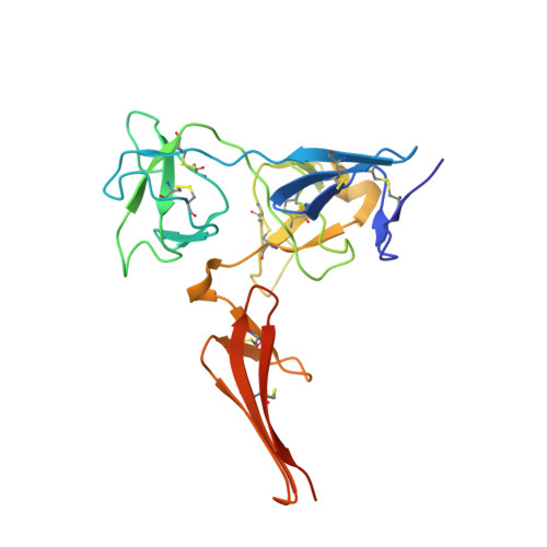
Implications for collagen binding from the crystallographic structure of fibronectin 6FnI1-2FnII7FnI

Erat, M.C.Schwarz-Linek, U.Pickford, A.R.Farndale, R.W.Campbell, I.D.Vakonakis, I.

(2010) J Biol Chem 285: 33764-33770

  • DOI: https://doi.org/10.1074/jbc.M110.139394
  • Primary Citation of Related Structures:  
    3MQL

  • PubMed Abstract: 

    Collagen and fibronectin (FN) are two abundant and essential components of the vertebrate extracellular matrix; they interact directly with cellular receptors and affect cell adhesion and migration. Past studies identified a FN fragment comprising six modules, (6)FnI(1-2)FnII(7-9)FnI, and termed the gelatin binding domain (GBD) as responsible for collagen interaction. Recently, we showed that the GBD binds tightly to a specific site within type I collagen and determined the structure of domains (8-9)FnI in complex with a peptide from that site. Here, we present the crystallographic structure of domains (6)FnI(1-2)FnII(7)FnI, which form a compact, globular unit through interdomain interactions. Analysis of NMR titrations with single-stranded collagen peptides reveals a dominant collagen interaction surface on domains (2)FnII and (7)FnI; a similar surface appears involved in interactions with triple-helical peptides. Models of the complete GBD, based on the new structure and the (8-9)FnI·collagen complex show a continuous putative collagen binding surface. We explore the implications of this model using long collagen peptides and discuss our findings in the context of FN interactions with collagen fibrils.


  • Organizational Affiliation

    Department of Biochemistry, University of Oxford, Oxford OX1 3QU, United Kingdom.


Macromolecules
Find similar proteins by:  (by identity cutoff)  |  3D Structure
Entity ID: 1
MoleculeChains Sequence LengthOrganismDetailsImage
Fibronectin 1220Homo sapiensMutation(s): 1 
Gene Names: FN1hCG_1813428
UniProt & NIH Common Fund Data Resources
Find proteins for P02751 (Homo sapiens)
Explore P02751 
Go to UniProtKB:  P02751
PHAROS:  P02751
GTEx:  ENSG00000115414 
Entity Groups  
Sequence Clusters30% Identity50% Identity70% Identity90% Identity95% Identity100% Identity
UniProt GroupP02751
Sequence Annotations
Expand
  • Reference Sequence
Experimental Data & Validation

Experimental Data

  • Method: X-RAY DIFFRACTION
  • Resolution: 3.00 Å
  • R-Value Free: 0.231 
  • R-Value Work: 0.201 
  • R-Value Observed: 0.203 
  • Space Group: I 41 2 2
Unit Cell:
Length ( Å )Angle ( ˚ )
a = 103.947α = 90
b = 103.947β = 90
c = 102.143γ = 90
Software Package:
Software NamePurpose
MAR345dtbdata collection
PHASERphasing
PHENIXrefinement
XSCALEdata scaling
SCALAdata scaling

Structure Validation

View Full Validation Report


Ligand Structure Quality Assessment 


Entry History 

Deposition Data

Revision History  (Full details and data files)

  • Version 1.0: 2010-08-25
    Type: Initial release
  • Version 1.1: 2011-07-13
    Changes: Version format compliance
  • Version 1.2: 2020-07-29
    Type: Remediation
    Reason: Carbohydrate remediation
    Changes: Data collection, Database references, Derived calculations, Structure summary
  • Version 1.3: 2023-11-01
    Changes: Data collection, Database references, Refinement description, Structure summary