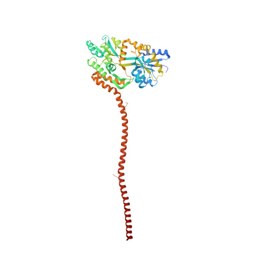
3MQ9

Crystal Structure of Ectodomain Mutant of BST-2/Tetherin/CD317 Fused to MBP


Experimental Data Snapshot

  • Method: X-RAY DIFFRACTION
  • Resolution: 2.80 Å
  • R-Value Free: 0.279 
  • R-Value Work: 0.231 
  • R-Value Observed: 0.233 

wwPDB Validation   3D Report Full Report


This is version 1.4 of the entry. See complete history


Literature

Structural insight into the mechanisms of enveloped virus tethering by tetherin.

Yang, H.Wang, J.Jia, X.McNatt, M.W.Zang, T.Pan, B.Meng, W.Wang, H.W.Bieniasz, P.D.Xiong, Y.

(2010) Proc Natl Acad Sci U S A 107: 18428-18432

  • DOI: https://doi.org/10.1073/pnas.1011485107
  • Primary Citation of Related Structures:  
    3MQ7, 3MQ9, 3MQB, 3MQC

  • PubMed Abstract: 

    Tetherin/BST2 is a type-II membrane protein that inhibits the release of a range of enveloped viruses, including HIV-1. Here we report three crystal structures of human tetherin, including the full-length ectodomain, a triple cysteine mutant and an ectodomain truncation. These structures show that tetherin forms a continuous alpha helix encompassing almost the entire ectodomain. Tetherin helices dimerize into parallel coiled coils via interactions throughout the C-terminal portion of the ectodomain. A comparison of the multiple structures of the tetherin dimer reveals inherent constrained flexibility at two hinges positioned at residues A88 and G109. In the crystals, two tetherin ectodomain dimers associate into a tetramer by forming an antiparallel four-helix bundle at their N termini. However, mutagenesis studies suggest that the tetrametric form of tetherin, although potentially contributing to, is not essential for its antiviral activity. Nonetheless, the structural and chemical properties of the N terminus of the ectodomain are important for optimal tethering function. This study provides detailed insight into the mechanisms by which this broad-spectrum antiviral restriction factor can function.


  • Organizational Affiliation

    Department of Molecular Biophysics and Biochemistry, Yale University, New Haven, CT 06520-8114, USA.


Macromolecules
Find similar proteins by:  (by identity cutoff)  |  3D Structure
Entity ID: 1
MoleculeChains Sequence LengthOrganismDetailsImage
Bone marrow stromal antigen 2 fused to Maltose-binding periplasmic protein
A, B, C, D, E
A, B, C, D, E, F, G, H
471Escherichia coli K-12Homo sapiens
This entity is chimeric
Mutation(s): 1 
Gene Names: malEb4034JW3994BST2
UniProt & NIH Common Fund Data Resources
Find proteins for P0AEX9 (Escherichia coli (strain K12))
Explore P0AEX9 
Go to UniProtKB:  P0AEX9
Find proteins for Q10589 (Homo sapiens)
Explore Q10589 
Go to UniProtKB:  Q10589
PHAROS:  Q10589
GTEx:  ENSG00000130303 
Entity Groups  
Sequence Clusters30% Identity50% Identity70% Identity90% Identity95% Identity100% Identity
UniProt GroupsQ10589P0AEX9
Sequence Annotations
Expand
  • Reference Sequence
Small Molecules
Modified Residues  1 Unique
IDChains TypeFormula2D DiagramParent
MSE
Query on MSE
A, B, C, D, E
A, B, C, D, E, F, G, H
L-PEPTIDE LINKINGC5 H11 N O2 SeMET
Experimental Data & Validation

Experimental Data

  • Method: X-RAY DIFFRACTION
  • Resolution: 2.80 Å
  • R-Value Free: 0.279 
  • R-Value Work: 0.231 
  • R-Value Observed: 0.233 
  • Space Group: P 1 21 1
Unit Cell:
Length ( Å )Angle ( ˚ )
a = 94.502α = 90
b = 202.443β = 90.41
c = 107.277γ = 90
Software Package:
Software NamePurpose
ADSCdata collection
PHASERphasing
REFMACrefinement
HKL-2000data reduction
HKL-2000data scaling

Structure Validation

View Full Validation Report


Entry History 

Deposition Data

Revision History  (Full details and data files)

  • Version 1.0: 2010-10-27
    Type: Initial release
  • Version 1.1: 2011-07-13
    Changes: Version format compliance
  • Version 1.2: 2017-08-16
    Changes: Refinement description, Source and taxonomy
  • Version 1.3: 2023-09-06
    Changes: Data collection, Database references, Derived calculations, Refinement description
  • Version 1.4: 2023-11-22
    Changes: Data collection