3MQ6

Domain swapped SgrAI with DNA and calcium bound


Experimental Data Snapshot

  • Method: X-RAY DIFFRACTION
  • Resolution: 2.00 Å
  • R-Value Free: 0.261 
  • R-Value Work: 0.211 
  • R-Value Observed: 0.212 

wwPDB Validation   3D Report Full Report


This is version 1.2 of the entry. See complete history


Literature

Domain swapping in allosteric modulation of DNA specificity.

Park, C.K.Joshi, H.K.Agrawal, A.Ghare, M.I.Little, E.J.Dunten, P.W.Bitinaite, J.Horton, N.C.

(2010) PLoS Biol 8: e1000554-e1000554

  • DOI: https://doi.org/10.1371/journal.pbio.1000554
  • Primary Citation of Related Structures:  
    3MQ6

  • PubMed Abstract: 

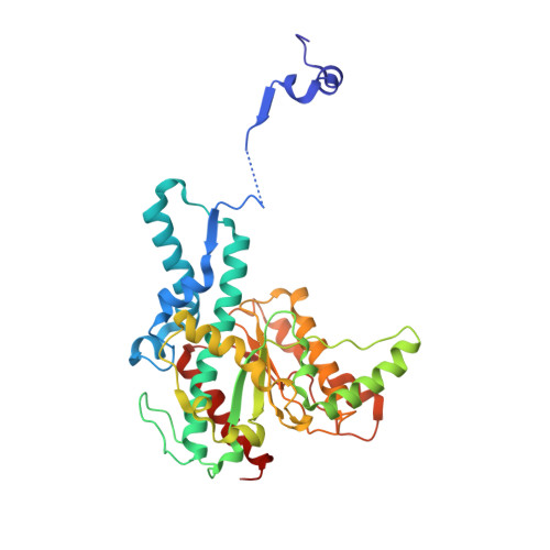
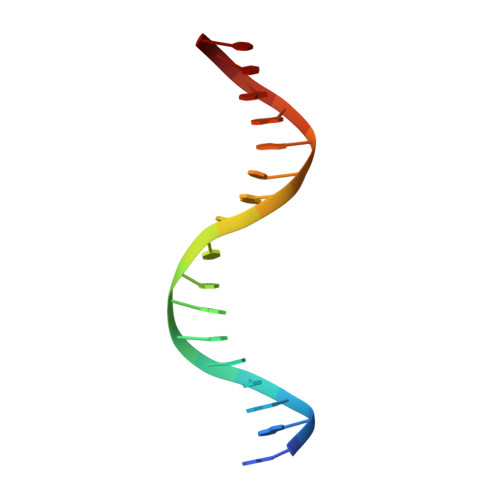
    SgrAI is a type IIF restriction endonuclease that cuts an unusually long recognition sequence and exhibits allosteric self-modulation of cleavage activity and sequence specificity. Previous studies have shown that DNA bound dimers of SgrAI oligomerize into an activated form with higher DNA cleavage rates, although previously determined crystal structures of SgrAI bound to DNA show only the DNA bound dimer. A new crystal structure of the type II restriction endonuclease SgrAI bound to DNA and Ca(2+) is now presented, which shows the close association of two DNA bound SgrAI dimers. This tetrameric form is unlike those of the homologous enzymes Cfr10I and NgoMIV and is formed by the swapping of the amino-terminal 24 amino acid residues. Two mutations predicted to destabilize the swapped form of SgrAI, P27W and P27G, have been made and shown to eliminate both the oligomerization of the DNA bound SgrAI dimers as well as the allosteric stimulation of DNA cleavage by SgrAI. A mechanism involving domain swapping is proposed to explain the unusual allosteric properties of SgrAI via association of the domain swapped tetramer of SgrAI bound to DNA into higher order oligomers.


  • Organizational Affiliation

    Department of Chemistry and Biochemistry, University of Arizona, Tucson, Arizona, United States of America.


Macromolecules

Find similar proteins by:  (by identity cutoff)  |  3D Structure
Entity ID: 1
MoleculeChains Sequence LengthOrganismDetailsImage
SgraIR restriction enzyme
A, B, C, D, E
A, B, C, D, E, F, G, H
338Streptomyces griseusMutation(s): 0 
Gene Names: sgraIR
EC: 3.1.21.4
UniProt
Find proteins for Q9F6L0 (Streptomyces griseus)
Explore Q9F6L0 
Go to UniProtKB:  Q9F6L0
Entity Groups  
Sequence Clusters30% Identity50% Identity70% Identity90% Identity95% Identity100% Identity
UniProt GroupQ9F6L0
Sequence Annotations
Expand
  • Reference Sequence

Find similar nucleic acids by:  Sequence   |   3D Structure  

Entity ID: 2
MoleculeChains LengthOrganismImage
DNA (5'-D(*AP*AP*GP*TP*CP*CP*AP*CP*CP*GP*GP*TP*GP*GP*AP*CP*T)-3')17N/A
Sequence Annotations
Expand
  • Reference Sequence
Small Molecules
Ligands 1 Unique
IDChains Name / Formula / InChI Key2D Diagram3D Interactions
CA
Query on CA

Download Ideal Coordinates CCD File 
AA [auth D]
BA [auth D]
CA [auth E]
DA [auth E]
EA [auth F]
AA [auth D],
BA [auth D],
CA [auth E],
DA [auth E],
EA [auth F],
FA [auth F],
GA [auth F],
HA [auth G],
IA [auth G],
JA [auth H],
KA [auth H],
LA [auth H],
Q [auth A],
R [auth A],
S [auth A],
T [auth B],
U [auth B],
V [auth B],
W [auth C],
X [auth C],
Y [auth C],
Z [auth D]
CALCIUM ION
Ca
BHPQYMZQTOCNFJ-UHFFFAOYSA-N
Experimental Data & Validation

Experimental Data

  • Method: X-RAY DIFFRACTION
  • Resolution: 2.00 Å
  • R-Value Free: 0.261 
  • R-Value Work: 0.211 
  • R-Value Observed: 0.212 
  • Space Group: P 21 21 21
Unit Cell:
Length ( Å )Angle ( ˚ )
a = 130.384α = 90
b = 134.949β = 90
c = 237.485γ = 90
Software Package:
Software NamePurpose
Blu-Icedata collection
PHENIXrefinement
HKL-2000data reduction
HKL-2000data scaling

Structure Validation

View Full Validation Report


Entry History 

Deposition Data

Revision History  (Full details and data files)

  • Version 1.0: 2010-11-10
    Type: Initial release
  • Version 1.1: 2011-07-13
    Changes: Version format compliance
  • Version 1.2: 2024-02-21
    Changes: Data collection, Database references, Derived calculations