3MOQ

Amyloid beta(18-41) peptide fusion with new antigen receptor variable domain from sharks


Experimental Data Snapshot

  • Method: X-RAY DIFFRACTION
  • Resolution: 2.05 Å
  • R-Value Free: 0.249 
  • R-Value Work: 0.190 
  • R-Value Observed: 0.193 

wwPDB Validation   3D Report Full Report


This is version 1.3 of the entry. See complete history


Literature

Crystal Structure of the Amyloid-{beta} p3 Fragment Provides a Model for Oligomer Formation in Alzheimer's Disease

Streltsov, V.A.Varghese, J.N.Masters, C.L.Nuttall, S.D.

(2011) J Neurosci 31: 1419-1426

  • DOI: https://doi.org/10.1523/JNEUROSCI.4259-10.2011
  • Primary Citation of Related Structures:  
    3MOQ

  • PubMed Abstract: 

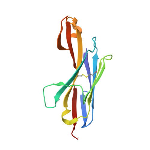
    Alzheimer's disease is a progressive neurodegenerative disorder associated with the presence of amyloid-β (Aβ) peptide fibrillar plaques in the brain. However, current evidence suggests that soluble nonfibrillar Aβ oligomers may be the major drivers of Aβ-mediated synaptic dysfunction. Structural information on these Aβ species has been very limited because of their noncrystalline and unstable nature. Here, we describe a crystal structure of amylogenic residues 18-41 of the Aβ peptide (equivalent to the p3 α/γ-secretase fragment of amyloid precursor protein) presented within the CDR3 loop region of a shark Ig new antigen receptor (IgNAR) single variable domain antibody. The predominant oligomeric species is a tightly associated Aβ dimer, with paired dimers forming a tetramer in the crystal caged within four IgNAR domains, preventing uncontrolled amyloid formation. Our structure correlates with independently observed features of small nonfibrillar Aβ oligomers and reveals conserved elements consistent with residues and motifs predicted as critical in Aβ folding and oligomerization, thus potentially providing a model system for nonfibrillar oligomer formation in Alzheimer's disease.


  • Organizational Affiliation

    Commonwealth Scientific and Industrial Research Organization, Parkville, Victoria 3052, Australia. victor.streltsov@csiro.au


Macromolecules
Find similar proteins by:  (by identity cutoff)  |  3D Structure
Entity ID: 1
MoleculeChains Sequence LengthOrganismDetailsImage
New antigen receptor variable domain,P3(40) peptide from Amyloid beta A4 protein,New antigen receptor variable domain
A, B, C, D
126Orectolobus maculatusHomo sapiensMutation(s): 0 
UniProt & NIH Common Fund Data Resources
Find proteins for Q6X1E6 (Orectolobus maculatus)
Explore Q6X1E6 
Go to UniProtKB:  Q6X1E6
Find proteins for P05067 (Homo sapiens)
Explore P05067 
Go to UniProtKB:  P05067
PHAROS:  P05067
GTEx:  ENSG00000142192 
Entity Groups  
Sequence Clusters30% Identity50% Identity70% Identity90% Identity95% Identity100% Identity
UniProt GroupsQ6X1E6P05067
Sequence Annotations
Expand
  • Reference Sequence
Experimental Data & Validation

Experimental Data

  • Method: X-RAY DIFFRACTION
  • Resolution: 2.05 Å
  • R-Value Free: 0.249 
  • R-Value Work: 0.190 
  • R-Value Observed: 0.193 
  • Space Group: P 32
Unit Cell:
Length ( Å )Angle ( ˚ )
a = 79.653α = 90
b = 79.653β = 90
c = 85.457γ = 120
Software Package:
Software NamePurpose
MOLREPphasing
REFMACrefinement
HKL-2000data reduction
HKL-2000data scaling

Structure Validation

View Full Validation Report


Entry History 

Deposition Data

Revision History  (Full details and data files)

  • Version 1.0: 2011-02-16
    Type: Initial release
  • Version 1.1: 2011-07-13
    Changes: Version format compliance
  • Version 1.2: 2017-06-21
    Changes: Database references, Source and taxonomy, Structure summary
  • Version 1.3: 2023-11-01
    Changes: Data collection, Database references, Refinement description