3MF0

Crystal structure of PDE5A GAF domain (89-518)


Experimental Data Snapshot

  • Method: X-RAY DIFFRACTION
  • Resolution: 3.10 Å
  • R-Value Free: 0.289 
  • R-Value Work: 0.232 
  • R-Value Observed: 0.235 

wwPDB Validation   3D Report Full Report


This is version 1.3 of the entry. See complete history


Literature

Conformation changes, N-terminal involvement, and cGMP signal relay in the phosphodiesterase-5 GAF domain.

Wang, H.Robinson, H.Ke, H.

(2010) J Biol Chem 285: 38149-38156

  • DOI: https://doi.org/10.1074/jbc.M110.141614
  • Primary Citation of Related Structures:  
    3LFV, 3MF0

  • PubMed Abstract: 

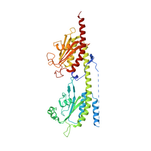
    The activity of phosphodiesterase-5 (PDE5) is specific for cGMP and is regulated by cGMP binding to GAF-A in its regulatory domain. To better understand the regulatory mechanism, x-ray crystallographic and biochemical studies were performed on constructs of human PDE5A1 containing the N-terminal phosphorylation segment, GAF-A, and GAF-B. Superposition of this unliganded GAF-A with the previously reported NMR structure of cGMP-bound PDE5 revealed dramatic conformational differences and suggested that helix H4 and strand B3 probably serve as two lids to gate the cGMP-binding pocket in GAF-A. The structure also identified an interfacial region among GAF-A, GAF-B, and the N-terminal loop, which may serve as a relay of the cGMP signal from GAF-A to GAF-B. N-terminal loop 98-147 was physically associated with GAF-B domains of the dimer. Biochemical analyses showed an inhibitory effect of this loop on cGMP binding and its involvement in the cGMP-induced conformation changes.


  • Organizational Affiliation

    Department of Biochemistry and Biophysics and Lineberger Comprehensive Cancer Center, University of North Carolina, Chapel Hill, North Carolina 27599-7260, USA.


Macromolecules
Find similar proteins by:  (by identity cutoff)  |  3D Structure
Entity ID: 1
MoleculeChains Sequence LengthOrganismDetailsImage
cGMP-specific 3',5'-cyclic phosphodiesterase
A, B
432Homo sapiensMutation(s): 1 
Gene Names: PDE5PDE5APDE5A1
EC: 3.1.4.35
UniProt & NIH Common Fund Data Resources
Find proteins for O76074 (Homo sapiens)
Explore O76074 
Go to UniProtKB:  O76074
PHAROS:  O76074
GTEx:  ENSG00000138735 
Entity Groups  
Sequence Clusters30% Identity50% Identity70% Identity90% Identity95% Identity100% Identity
UniProt GroupO76074
Sequence Annotations
Expand
  • Reference Sequence
Experimental Data & Validation

Experimental Data

  • Method: X-RAY DIFFRACTION
  • Resolution: 3.10 Å
  • R-Value Free: 0.289 
  • R-Value Work: 0.232 
  • R-Value Observed: 0.235 
  • Space Group: P 31 2 1
Unit Cell:
Length ( Å )Angle ( ˚ )
a = 144.534α = 90
b = 144.534β = 90
c = 135.008γ = 120
Software Package:
Software NamePurpose
ADSCdata collection
AMoREphasing
REFMACrefinement
HKL-2000data reduction
HKL-2000data scaling

Structure Validation

View Full Validation Report


Entry History 

Deposition Data

Revision History  (Full details and data files)

  • Version 1.0: 2010-09-22
    Type: Initial release
  • Version 1.1: 2011-07-13
    Changes: Version format compliance
  • Version 1.2: 2021-10-06
    Changes: Database references
  • Version 1.3: 2024-02-21
    Changes: Data collection