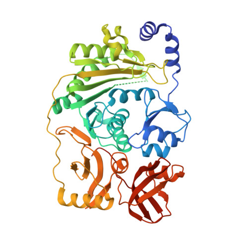
Multi-site-specific 16S rRNA methyltransferase RsmF from Thermus thermophilus.
Demirci, H., Larsen, L.H., Hansen, T., Rasmussen, A., Cadambi, A., Gregory, S.T., Kirpekar, F., Jogl, G.(2010) RNA 16: 1584-1596
- PubMed: 20558545
- DOI: https://doi.org/10.1261/rna.2088310
- Primary Citation of Related Structures:
3M6U, 3M6V, 3M6W, 3M6X - PubMed Abstract:
Cells devote a significant effort toward the production of multiple modified nucleotides in rRNAs, which fine tune the ribosome function. Here, we report that two methyltransferases, RsmB and RsmF, are responsible for all four 5-methylcytidine (m(5)C) modifications in 16S rRNA of Thermus thermophilus. Like Escherichia coli RsmB, T. thermophilus RsmB produces m(5)C967. In contrast to E. coli RsmF, which introduces a single m(5)C1407 modification, T. thermophilus RsmF modifies three positions, generating m(5)C1400 and m(5)C1404 in addition to m(5)C1407. These three residues are clustered near the decoding site of the ribosome, but are situated in distinct structural contexts, suggesting a requirement for flexibility in the RsmF active site that is absent from the E. coli enzyme. Two of these residues, C1400 and C1404, are sufficiently buried in the mature ribosome structure so as to require extensive unfolding of the rRNA to be accessible to RsmF. In vitro, T. thermophilus RsmF methylates C1400, C1404, and C1407 in a 30S subunit substrate, but only C1400 and C1404 when naked 16S rRNA is the substrate. The multispecificity of T. thermophilus RsmF is potentially explained by three crystal structures of the enzyme in a complex with cofactor S-adenosyl-methionine at up to 1.3 A resolution. In addition to confirming the overall structural similarity to E. coli RsmF, these structures also reveal that key segments in the active site are likely to be dynamic in solution, thereby expanding substrate recognition by T. thermophilus RsmF.
Organizational Affiliation:
Department of Molecular Biology, Cell Biology and Biochemistry, Brown University, Providence, RI 02912, USA.