3M4F

Structural insights into the acidophilic pH adaptation of a novel endo-1,4-beta-xylanase from Scytalidium acidophilum


Experimental Data Snapshot

  • Method: X-RAY DIFFRACTION
  • Resolution: 1.89 Å
  • R-Value Free: 0.197 
  • R-Value Work: 0.167 
  • R-Value Observed: 0.168 

wwPDB Validation   3D Report Full Report


Ligand Structure Quality Assessment 


This is version 1.2 of the entry. See complete history


Literature

Structural insights into the acidophilic pH adaptation of a novel endo-1,4-beta-xylanase from Scytalidium acidophilum

Michaux, C.Pouyez, J.Mayard, A.Vandurm, P.Housen, I.Wouters, J.

(2010) Biochimie 92: 1407-1415

  • DOI: https://doi.org/10.1016/j.biochi.2010.07.003
  • Primary Citation of Related Structures:  
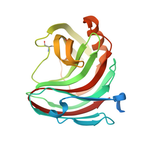
    3M4F

  • PubMed Abstract: 

    In this study, the crystal structure of a novel endo-1,4-β-xylanase from Scytalidium acidophilum, XYL1, was solved at 1.9Å resolution. This is one of the few solved crystal structures of acidophilic proteins. The enzyme has the overall fold typical to family 11 xylanases. Comparison of this structure with other homologous acidophilic, neutrophilic and alkalophilic xylanases provides additional insights into the general features involved in low pH adaptation (stability and activity). Several sequence and structure modifications appeared to be responsible for the acidophilic characteristic: (a) the presence of an aspartic acid H bonded to the acid/base catalyst (b) the nature of specifically conserved residues in the active site (c) the negative potential at the surface (d) the decreased number of salt bridges and H bonds in comparison with highly alkaline enzymes.


  • Organizational Affiliation

    Unité de Chimie Physique Structurale et Théorique, Chemistry Department, FUNDP, 61 rue de Bruxelles, B-5000 Namur, Belgium. catherine.michaux@fundp.ac.be


Macromolecules
Find similar proteins by:  (by identity cutoff)  |  3D Structure
Entity ID: 1
MoleculeChains Sequence LengthOrganismDetailsImage
Endo-1,4-beta-xylanase
A, B, C, D
205Acidomyces acidophilusMutation(s): 0 
Gene Names: Xyl1
EC: 3.2.1.8
UniProt
Find proteins for Q6VAY1 (Acidomyces acidophilus)
Explore Q6VAY1 
Go to UniProtKB:  Q6VAY1
Entity Groups  
Sequence Clusters30% Identity50% Identity70% Identity90% Identity95% Identity100% Identity
UniProt GroupQ6VAY1
Sequence Annotations
Expand
  • Reference Sequence
Experimental Data & Validation

Experimental Data

  • Method: X-RAY DIFFRACTION
  • Resolution: 1.89 Å
  • R-Value Free: 0.197 
  • R-Value Work: 0.167 
  • R-Value Observed: 0.168 
  • Space Group: P 21 3
Unit Cell:
Length ( Å )Angle ( ˚ )
a = 144.66α = 90
b = 144.66β = 90
c = 144.66γ = 90
Software Package:
Software NamePurpose
ADSCdata collection
PHASESphasing
REFMACrefinement
XDSdata reduction
XSCALEdata scaling

Structure Validation

View Full Validation Report


Ligand Structure Quality Assessment 


Entry History 

Deposition Data

Revision History  (Full details and data files)

  • Version 1.0: 2010-07-28
    Type: Initial release
  • Version 1.1: 2011-07-13
    Changes: Version format compliance
  • Version 1.2: 2023-11-01
    Changes: Data collection, Database references, Derived calculations, Refinement description