3LVK

Crystal Structure of E.coli IscS-TusA complex (form 2)


Experimental Data Snapshot

  • Method: X-RAY DIFFRACTION
  • Resolution: 2.44 Å
  • R-Value Free: 0.249 
  • R-Value Work: 0.207 
  • R-Value Observed: 0.209 

wwPDB Validation   3D Report Full Report


Ligand Structure Quality Assessment 


This is version 1.3 of the entry. See complete history


Literature

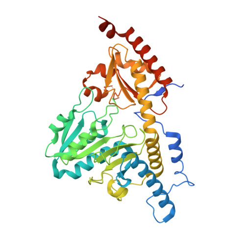
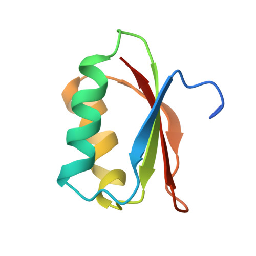
Structural basis for Fe-S cluster assembly and tRNA thiolation mediated by IscS protein-protein interactions.

Shi, R.Proteau, A.Villarroya, M.Moukadiri, I.Zhang, L.Trempe, J.F.Matte, A.Armengod, M.E.Cygler, M.

(2010) PLoS Biol 8: e1000354-e1000354

  • DOI: https://doi.org/10.1371/journal.pbio.1000354
  • Primary Citation of Related Structures:  
    3LVJ, 3LVK, 3LVL, 3LVM

  • PubMed Abstract: 

    The cysteine desulfurase IscS is a highly conserved master enzyme initiating sulfur transfer via persulfide to a range of acceptor proteins involved in Fe-S cluster assembly, tRNA modifications, and sulfur-containing cofactor biosynthesis. Several IscS-interacting partners including IscU, a scaffold for Fe-S cluster assembly; TusA, the first member of a sulfur relay leading to sulfur incorporation into the wobble uridine of several tRNAs; ThiI, involved in tRNA modification and thiamine biosynthesis; and rhodanese RhdA are sulfur acceptors. Other proteins, such as CyaY/frataxin and IscX, also bind to IscS, but their functional roles are not directly related to sulfur transfer. We have determined the crystal structures of IscS-IscU and IscS-TusA complexes providing the first insight into their different modes of binding and the mechanism of sulfur transfer. Exhaustive mutational analysis of the IscS surface allowed us to map the binding sites of various partner proteins and to determine the functional and biochemical role of selected IscS and TusA residues. IscS interacts with its partners through an extensive surface area centered on the active site Cys328. The structures indicate that the acceptor proteins approach Cys328 from different directions and suggest that the conformational plasticity of a long loop containing this cysteine is essential for the ability of IscS to transfer sulfur to multiple acceptor proteins. The sulfur acceptors can only bind to IscS one at a time, while frataxin and IscX can form a ternary complex with IscU and IscS. Our data support the role of frataxin as an iron donor for IscU to form the Fe-S clusters.


  • Organizational Affiliation

    Department of Biochemistry, McGill University, Montréal, Québec, Canada.


Macromolecules
Find similar proteins by:  (by identity cutoff)  |  3D Structure
Entity ID: 1
MoleculeChains Sequence LengthOrganismDetailsImage
Cysteine desulfurase423Escherichia coli O157:H7 str. EDL933Mutation(s): 0 
Gene Names: iscS
EC: 2.8.1.7
UniProt
Find proteins for P0A6B9 (Escherichia coli O157:H7)
Explore P0A6B9 
Go to UniProtKB:  P0A6B9
Entity Groups  
Sequence Clusters30% Identity50% Identity70% Identity90% Identity95% Identity100% Identity
UniProt GroupP0A6B9
Sequence Annotations
Expand
  • Reference Sequence
Find similar proteins by:  (by identity cutoff)  |  3D Structure
Entity ID: 2
MoleculeChains Sequence LengthOrganismDetailsImage
Sulfurtransferase tusA82Escherichia coli O157:H7 str. EDL933Mutation(s): 0 
Gene Names: tusA
EC: 2.8.1
UniProt
Find proteins for P0A892 (Escherichia coli O157:H7)
Explore P0A892 
Go to UniProtKB:  P0A892
Entity Groups  
Sequence Clusters30% Identity50% Identity70% Identity90% Identity95% Identity100% Identity
UniProt GroupP0A892
Sequence Annotations
Expand
  • Reference Sequence
Small Molecules
Ligands 1 Unique
IDChains Name / Formula / InChI Key2D Diagram3D Interactions
PLP
Query on PLP

Download Ideal Coordinates CCD File 
C [auth A]PYRIDOXAL-5'-PHOSPHATE
C8 H10 N O6 P
NGVDGCNFYWLIFO-UHFFFAOYSA-N
Experimental Data & Validation

Experimental Data

  • Method: X-RAY DIFFRACTION
  • Resolution: 2.44 Å
  • R-Value Free: 0.249 
  • R-Value Work: 0.207 
  • R-Value Observed: 0.209 
  • Space Group: C 2 2 21
Unit Cell:
Length ( Å )Angle ( ˚ )
a = 72.893α = 90
b = 131.407β = 90
c = 106.411γ = 90
Software Package:
Software NamePurpose
DENZOdata reduction
SCALEPACKdata scaling
PHENIXrefinement
PDB_EXTRACTdata extraction
HKL-2000data reduction
HKL-2000data scaling
PHASERphasing

Structure Validation

View Full Validation Report


Ligand Structure Quality Assessment 


Entry History 

Deposition Data

Revision History  (Full details and data files)

  • Version 1.0: 2010-04-21
    Type: Initial release
  • Version 1.1: 2011-07-13
    Changes: Version format compliance
  • Version 1.2: 2017-11-08
    Changes: Refinement description
  • Version 1.3: 2023-09-06
    Changes: Data collection, Database references, Derived calculations, Refinement description