3LOG

Crystal structure of MbtI from Mycobacterium tuberculosis


Experimental Data Snapshot

  • Method: X-RAY DIFFRACTION
  • Resolution: 1.73 Å
  • R-Value Free: 0.225 
  • R-Value Work: 0.181 
  • R-Value Observed: 0.183 

wwPDB Validation   3D Report Full Report


This is version 1.3 of the entry. See complete history


Literature

Inhibition studies of Mycobacterium tuberculosis salicylate synthase (MbtI).

Manos-Turvey, A.Bulloch, E.M.Rutledge, P.J.Baker, E.N.Lott, J.S.Payne, R.J.

(2010) ChemMedChem 5: 1067-1079

  • DOI: https://doi.org/10.1002/cmdc.201000137
  • Primary Citation of Related Structures:  
    3LOG

  • PubMed Abstract: 

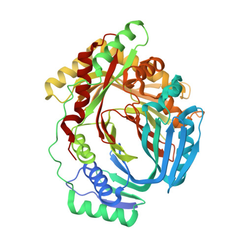
    Mycobacterium tuberculosis salicylate synthase (MbtI), a member of the chorismate-utilizing enzyme family, catalyses the first committed step in the biosynthesis of the siderophore mycobactin T. This complex secondary metabolite is essential for both virulence and survival of M. tuberculosis, the etiological agent of tuberculosis (TB). It is therefore anticipated that inhibitors of this enzyme may serve as TB therapies with a novel mode of action. Herein we describe the first inhibition study of M. tuberculosis MbtI using a library of functionalized benzoate-based inhibitors designed to mimic the substrate (chorismate) and intermediate (isochorismate) of the MbtI-catalyzed reaction. The most potent inhibitors prepared were those designed to mimic the enzyme intermediate, isochorismate. These compounds, based on a 2,3-dihydroxybenzoate scaffold, proved to be low-micromolar inhibitors of MbtI. The most potent inhibitors in this series possessed hydrophobic enol ether side chains at C3 in place of the enol-pyruvyl side chain found in chorismate and isochorismate.


  • Organizational Affiliation

    School of Chemistry, The University of Sydney, NSW 2006, Australia.


Macromolecules
Find similar proteins by:  (by identity cutoff)  |  3D Structure
Entity ID: 1
MoleculeChains Sequence LengthOrganismDetailsImage
Isochorismate synthase/isochorismate-pyruvate lyase mbtI
A, B, C, D
451Mycobacterium tuberculosisMutation(s): 0 
Gene Names: mbtIMT2454Rv2386c
EC: 5.4.4.2 (PDB Primary Data), 4.1.3 (PDB Primary Data)
UniProt
Find proteins for P9WFX1 (Mycobacterium tuberculosis (strain ATCC 25618 / H37Rv))
Explore P9WFX1 
Go to UniProtKB:  P9WFX1
Entity Groups  
Sequence Clusters30% Identity50% Identity70% Identity90% Identity95% Identity100% Identity
UniProt GroupP9WFX1
Sequence Annotations
Expand
  • Reference Sequence
Small Molecules
Ligands 4 Unique
IDChains Name / Formula / InChI Key2D Diagram3D Interactions
SIN
Query on SIN

Download Ideal Coordinates CCD File 
E [auth A],
F [auth A],
H [auth B],
L [auth C],
O [auth D]
SUCCINIC ACID
C4 H6 O4
KDYFGRWQOYBRFD-UHFFFAOYSA-N
GOL
Query on GOL

Download Ideal Coordinates CCD File 
G [auth A],
K [auth B],
M [auth C],
N [auth C],
P [auth D]
GLYCEROL
C3 H8 O3
PEDCQBHIVMGVHV-UHFFFAOYSA-N
CO3
Query on CO3

Download Ideal Coordinates CCD File 
J [auth B]CARBONATE ION
C O3
BVKZGUZCCUSVTD-UHFFFAOYSA-L
NA
Query on NA

Download Ideal Coordinates CCD File 
I [auth B]SODIUM ION
Na
FKNQFGJONOIPTF-UHFFFAOYSA-N
Experimental Data & Validation

Experimental Data

  • Method: X-RAY DIFFRACTION
  • Resolution: 1.73 Å
  • R-Value Free: 0.225 
  • R-Value Work: 0.181 
  • R-Value Observed: 0.183 
  • Space Group: P 1 21 1
Unit Cell:
Length ( Å )Angle ( ˚ )
a = 87.87α = 90
b = 115.703β = 91.6
c = 93.893γ = 90
Software Package:
Software NamePurpose
DENZOdata reduction
SCALEPACKdata scaling
MOLREPphasing
REFMACrefinement
PDB_EXTRACTdata extraction
MAR345dtbdata collection
HKL-2000data reduction

Structure Validation

View Full Validation Report


Entry History 

Deposition Data

Revision History  (Full details and data files)

  • Version 1.0: 2011-02-09
    Type: Initial release
  • Version 1.1: 2011-07-13
    Changes: Version format compliance
  • Version 1.2: 2017-11-01
    Changes: Refinement description
  • Version 1.3: 2023-11-01
    Changes: Data collection, Database references, Derived calculations, Refinement description