3LGB

Crystal Structure of the Fe-S Domain of the yeast DNA primase


Experimental Data Snapshot

  • Method: X-RAY DIFFRACTION
  • Resolution: 1.54 Å
  • R-Value Free: 0.164 
  • R-Value Work: 0.154 
  • R-Value Observed: 0.155 

wwPDB Validation   3D Report Full Report


This is version 1.2 of the entry. See complete history


Literature

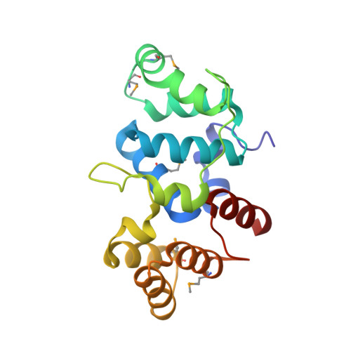
Shared Active Site Architecture between the Large Subunit of Eukaryotic Primase and DNA Photolyase

Sauguet, L.Klinge, S.Perera, R.L.Maman, J.D.Pellegrini, L.

(2010) PLoS One 5: 10083-10083

  • DOI: https://doi.org/10.1371/journal.pone.0010083
  • Primary Citation of Related Structures:  
    3LGB

  • PubMed Abstract: 

    DNA synthesis during replication relies on RNA primers synthesised by the primase, a specialised DNA-dependent RNA polymerase that can initiate nucleic acid synthesis de novo. In archaeal and eukaryotic organisms, the primase is a heterodimeric enzyme resulting from the constitutive association of a small (PriS) and large (PriL) subunit. The ability of the primase to initiate synthesis of an RNA primer depends on a conserved Fe-S domain at the C-terminus of PriL (PriL-CTD). However, the critical role of the PriL-CTD in the catalytic mechanism of initiation is not understood.


  • Organizational Affiliation

    Department of Biochemistry, University of Cambridge, Cambridge, United Kingdom.


Macromolecules
Find similar proteins by:  (by identity cutoff)  |  3D Structure
Entity ID: 1
MoleculeChains Sequence LengthOrganismDetailsImage
DNA primase large subunit
A, B
194Saccharomyces cerevisiaeMutation(s): 0 
Gene Names: PRI2YKL045WYKL258
EC: 2.7.7
UniProt
Find proteins for P20457 (Saccharomyces cerevisiae (strain ATCC 204508 / S288c))
Explore P20457 
Go to UniProtKB:  P20457
Entity Groups  
Sequence Clusters30% Identity50% Identity70% Identity90% Identity95% Identity100% Identity
UniProt GroupP20457
Sequence Annotations
Expand
  • Reference Sequence
Small Molecules
Ligands 4 Unique
IDChains Name / Formula / InChI Key2D Diagram3D Interactions
SF4
Query on SF4

Download Ideal Coordinates CCD File 
C [auth A],
F [auth B]
IRON/SULFUR CLUSTER
Fe4 S4
LJBDFODJNLIPKO-UHFFFAOYSA-N
EPE
Query on EPE

Download Ideal Coordinates CCD File 
D [auth A],
E [auth A]
4-(2-HYDROXYETHYL)-1-PIPERAZINE ETHANESULFONIC ACID
C8 H18 N2 O4 S
JKMHFZQWWAIEOD-UHFFFAOYSA-N
GOL
Query on GOL

Download Ideal Coordinates CCD File 
I [auth B]GLYCEROL
C3 H8 O3
PEDCQBHIVMGVHV-UHFFFAOYSA-N
ZN
Query on ZN

Download Ideal Coordinates CCD File 
G [auth B],
H [auth B]
ZINC ION
Zn
PTFCDOFLOPIGGS-UHFFFAOYSA-N
Modified Residues  1 Unique
IDChains TypeFormula2D DiagramParent
MSE
Query on MSE
A, B
L-PEPTIDE LINKINGC5 H11 N O2 SeMET
Experimental Data & Validation

Experimental Data

  • Method: X-RAY DIFFRACTION
  • Resolution: 1.54 Å
  • R-Value Free: 0.164 
  • R-Value Work: 0.154 
  • R-Value Observed: 0.155 
  • Space Group: P 61
Unit Cell:
Length ( Å )Angle ( ˚ )
a = 86.57α = 90
b = 86.57β = 90
c = 141.48γ = 120
Software Package:
Software NamePurpose
SOLVEphasing
BUSTERrefinement
MOSFLMdata reduction
SCALAdata scaling

Structure Validation

View Full Validation Report


Entry History 

Deposition Data

Revision History  (Full details and data files)

  • Version 1.0: 2010-04-21
    Type: Initial release
  • Version 1.1: 2011-07-13
    Changes: Version format compliance
  • Version 1.2: 2011-10-05
    Changes: Experimental preparation