


1.4A Crystal Structure of Isocitrate Lyase from Yersinia pestis CO92
Sharma, S.S., Brunzelle, J.S., Wawrzak, Z., Skarina, T., Gordon, E., Savchenko, A., Anderson, W.F.To be published.
Experimental Data Snapshot
wwPDB Validation 3D Report Full Report

Entity ID: 1 | |||||
|---|---|---|---|---|---|
| Molecule | Chains | Sequence Length | Organism | Details | Image |
| Isocitrate lyase | 435 | Yersinia pestis | Mutation(s): 0 Gene Names: aceA, y0016, YPO3725, YP_3087 EC: 4.1.3.1 | 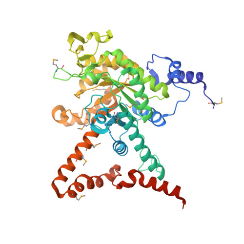 | |
UniProt | |||||
Find proteins for A0A2S9PCG6 (Yersinia pestis) Explore A0A2S9PCG6 Go to UniProtKB: A0A2S9PCG6 | |||||
Entity Groups | |||||
| Sequence Clusters | 30% Identity50% Identity70% Identity90% Identity95% Identity100% Identity | ||||
| UniProt Group | A0A2S9PCG6 | ||||
Sequence AnnotationsExpand | |||||
| |||||
| Modified Residues 1 Unique | |||||
|---|---|---|---|---|---|
| ID | Chains | Type | Formula | 2D Diagram | Parent |
| MSE Query on MSE | A, B | L-PEPTIDE LINKING | C5 H11 N O2 Se |  | MET |
| Length ( Å ) | Angle ( ˚ ) |
|---|---|
| a = 94.163 | α = 90 |
| b = 115.133 | β = 90 |
| c = 174.835 | γ = 90 |
| Software Name | Purpose |
|---|---|
| BLU-MAX | data collection |
| CRANK | phasing |
| ARP/wARP | model building |
| PHENIX | refinement |
| HKL-2000 | data reduction |
| HKL-2000 | data scaling |
RCSB PDB (citation) is hosted by
RCSB PDB is a member of the

