3LDW

Crystal Structure of Plasmodium vivax geranylgeranylpyrophosphate synthase PVX_092040 with zoledronate and IPP bound


Experimental Data Snapshot

  • Method: X-RAY DIFFRACTION
  • Resolution: 2.47 Å
  • R-Value Free: 0.286 
  • R-Value Work: 0.239 
  • R-Value Observed: 0.241 

wwPDB Validation   3D Report Full Report


Ligand Structure Quality Assessment 


This is version 1.2 of the entry. See complete history


Literature

Molecular characterization of a novel geranylgeranyl pyrophosphate synthase from Plasmodium parasites.

Artz, J.D.Wernimont, A.K.Dunford, J.E.Schapira, M.Dong, A.Zhao, Y.Lew, J.Russell, R.G.Ebetino, F.H.Oppermann, U.Hui, R.

(2011) J Biol Chem 286: 3315-3322

  • DOI: https://doi.org/10.1074/jbc.M109.027235
  • Primary Citation of Related Structures:  
    3LDW, 3MAV, 3PH7

  • PubMed Abstract: 

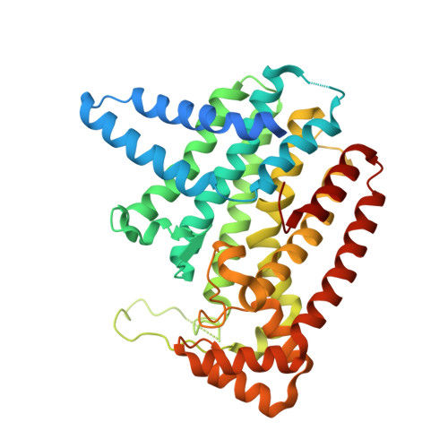
    We present here a study of a eukaryotic trans-prenylsynthase from the malaria pathogen Plasmodium vivax. Based on the results of biochemical assays and contrary to previous indications, this enzyme catalyzes the production of geranylgeranyl pyrophosphate (GGPP) rather than farnesyl pyrophosphate (FPP). Structural analysis shows that the product length is constrained by a hydrophobic cavity formed primarily by a set of residues from the same subunit as the product as well as at least one other from the dimeric partner. Furthermore, Plasmodium GGPP synthase (GGPPS) can bind nitrogen-containing bisphosphonates (N-BPs) strongly with the energetically favorable cooperation of three Mg(2+), resulting in inhibition by this class of compounds at IC(50) concentrations below 100 nM. In contrast, human and yeast GGPPSs do not accommodate a third magnesium atom in the same manner, resulting in their insusceptibility to N-BPs. This differentiation is in part attributable to a deviation in a conserved motif known as the second aspartate-rich motif: whereas the aspartates at the start and end of the five-residue motif in FFPP synthases and P. vivax GGPPSs both participate in the coordination of the third Mg(2+), an asparagine is featured as the last residue in human and yeast GGPPSs, resulting in a different manner of interaction with nitrogen-containing ligands.


  • Organizational Affiliation

    Structural Genomics Consortium, University of Toronto, Toronto, Ontario M5G 1L7, Canada.


Macromolecules
Find similar proteins by:  (by identity cutoff)  |  3D Structure
Entity ID: 1
MoleculeChains Sequence LengthOrganismDetailsImage
Farnesyl pyrophosphate synthase
A, B, C, D
396Plasmodium vivax Sal-1Mutation(s): 0 
Gene Names: PVX_092040
UniProt
Find proteins for A5K4U6 (Plasmodium vivax (strain Salvador I))
Explore A5K4U6 
Go to UniProtKB:  A5K4U6
Entity Groups  
Sequence Clusters30% Identity50% Identity70% Identity90% Identity95% Identity100% Identity
UniProt GroupA5K4U6
Sequence Annotations
Expand
  • Reference Sequence
Small Molecules
Ligands 5 Unique
IDChains Name / Formula / InChI Key2D Diagram3D Interactions
ZOL
Query on ZOL

Download Ideal Coordinates CCD File 
E [auth A],
L [auth B],
S [auth C],
X [auth D]
ZOLEDRONIC ACID
C5 H10 N2 O7 P2
XRASPMIURGNCCH-UHFFFAOYSA-N
IPE
Query on IPE

Download Ideal Coordinates CCD File 
F [auth A],
M [auth B],
T [auth C],
Y [auth D]
3-METHYLBUT-3-ENYL TRIHYDROGEN DIPHOSPHATE
C5 H12 O7 P2
NUHSROFQTUXZQQ-UHFFFAOYSA-N
GOL
Query on GOL

Download Ideal Coordinates CCD File 
CA [auth D],
K [auth A],
Q [auth B]
GLYCEROL
C3 H8 O3
PEDCQBHIVMGVHV-UHFFFAOYSA-N
EDO
Query on EDO

Download Ideal Coordinates CCD File 
J [auth A],
R [auth B]
1,2-ETHANEDIOL
C2 H6 O2
LYCAIKOWRPUZTN-UHFFFAOYSA-N
MG
Query on MG

Download Ideal Coordinates CCD File 
AA [auth D]
BA [auth D]
G [auth A]
H [auth A]
I [auth A]
AA [auth D],
BA [auth D],
G [auth A],
H [auth A],
I [auth A],
N [auth B],
O [auth B],
P [auth B],
U [auth C],
V [auth C],
W [auth C],
Z [auth D]
MAGNESIUM ION
Mg
JLVVSXFLKOJNIY-UHFFFAOYSA-N
Binding Affinity Annotations 
IDSourceBinding Affinity
ZOL PDBBind:  3LDW Ki: 10.7 (nM) from 1 assay(s)
Binding MOAD:  3LDW Ki: 10.7 (nM) from 1 assay(s)
Experimental Data & Validation

Experimental Data

  • Method: X-RAY DIFFRACTION
  • Resolution: 2.47 Å
  • R-Value Free: 0.286 
  • R-Value Work: 0.239 
  • R-Value Observed: 0.241 
  • Space Group: P 21 21 21
Unit Cell:
Length ( Å )Angle ( ˚ )
a = 111.264α = 90
b = 139.543β = 90
c = 109.711γ = 90
Software Package:
Software NamePurpose
CrystalCleardata collection
PHASERphasing
BUSTERrefinement
HKL-2000data reduction
HKL-2000data scaling

Structure Validation

View Full Validation Report


Ligand Structure Quality Assessment 


Entry History 

Revision History  (Full details and data files)

  • Version 1.0: 2010-11-17
    Type: Initial release
  • Version 1.1: 2011-07-13
    Changes: Version format compliance
  • Version 1.2: 2024-02-21
    Changes: Data collection, Database references, Derived calculations