3LBJ

Structure of human MDMX protein in complex with a small molecule inhibitor


Experimental Data Snapshot

  • Method: X-RAY DIFFRACTION
  • Resolution: 1.50 Å
  • R-Value Free: 0.206 
  • R-Value Work: 0.174 
  • R-Value Observed: 0.176 

wwPDB Validation   3D Report Full Report


Ligand Structure Quality Assessment 


This is version 1.4 of the entry. See complete history


Literature

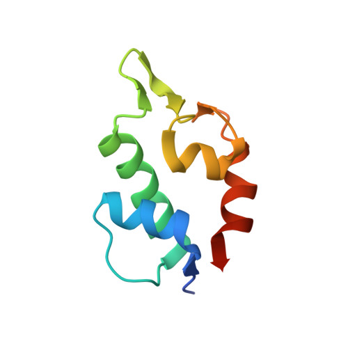
Structures of low molecular weight inhibitors bound to MDMX and MDM2 reveal new approaches for p53-MDMX/MDM2 antagonist drug discovery

Popowicz, G.M.Czarna, A.Wolf, S.Wang, K.Wang, W.Domling, A.Holak, T.A.

(2010) Cell Cycle 9: 1104-1111

  • DOI: https://doi.org/10.4161/cc.9.6.10956
  • Primary Citation of Related Structures:  
    3LBJ, 3LBK, 3LBL

  • PubMed Abstract: 

    Intensive anticancer drug discovery efforts have been made to develop small molecule inhibitors of the p53-MDM2 and p53-MDMX interactions. We present here the structures of the most potent inhibitors bound to MDM2 and MDMX that are based on the new imidazo-indole scaffold. In addition, the structure of the recently reported spiro-oxindole inhibitor bound to MDM2 is described. The structures indicate how the substituents of a small molecule that bind to the three subpockets of the MDM2/X-p53 interaction should be optimized for effective binding to MDM2 and/or MDMX. While the spiro-oxindole inhibitor triggers significant ligand-induced changes in MDM2, the imidazo-indoles share similar binding modes for MDMX and MDM2, but cause only minimal induced-fit changes in the structures of both proteins. Our study includes the first structure of the complex between MDMX and a small molecule and should aid in developing efficient scaffolds for binding to MDMX and/or MDM2.


  • Organizational Affiliation

    Max-Planck-Institute of Biochemistry, Martinsried, Germany.


Macromolecules
Find similar proteins by:  (by identity cutoff)  |  3D Structure
Entity ID: 1
MoleculeChains Sequence LengthOrganismDetailsImage
Protein Mdm4A [auth E]90Homo sapiensMutation(s): 0 
Gene Names: MDM4MDMX
UniProt & NIH Common Fund Data Resources
Find proteins for O15151 (Homo sapiens)
Explore O15151 
Go to UniProtKB:  O15151
PHAROS:  O15151
GTEx:  ENSG00000198625 
Entity Groups  
Sequence Clusters30% Identity50% Identity70% Identity90% Identity95% Identity100% Identity
UniProt GroupO15151
Sequence Annotations
Expand
  • Reference Sequence
Small Molecules
Ligands 2 Unique
IDChains Name / Formula / InChI Key2D Diagram3D Interactions
WW8
Query on WW8

Download Ideal Coordinates CCD File 
B [auth E],
C [auth E]
N-[(3S)-1-({6-chloro-3-[1-(4-chlorobenzyl)-4-phenyl-1H-imidazol-5-yl]-1H-indol-2-yl}carbonyl)pyrrolidin-3-yl]-N,N',N'-trimethylpropane-1,3-diamine
C35 H38 Cl2 N6 O
LWWQYOWAPGRPRH-NDEPHWFRSA-N
SO4
Query on SO4

Download Ideal Coordinates CCD File 
D [auth E]SULFATE ION
O4 S
QAOWNCQODCNURD-UHFFFAOYSA-L
Binding Affinity Annotations 
IDSourceBinding Affinity
WW8 PDBBind:  3LBJ Ki: 1.10e+4 (nM) from 1 assay(s)
Experimental Data & Validation

Experimental Data

  • Method: X-RAY DIFFRACTION
  • Resolution: 1.50 Å
  • R-Value Free: 0.206 
  • R-Value Work: 0.174 
  • R-Value Observed: 0.176 
  • Space Group: I 21 3
Unit Cell:
Length ( Å )Angle ( ˚ )
a = 80.97α = 90
b = 80.97β = 90
c = 80.97γ = 90
Software Package:
Software NamePurpose
MAR345data collection
MOLREPphasing
REFMACrefinement
XDSdata reduction
XSCALEdata scaling

Structure Validation

View Full Validation Report


Ligand Structure Quality Assessment 


Entry History 

Deposition Data

Revision History  (Full details and data files)

  • Version 1.0: 2010-03-16
    Type: Initial release
  • Version 1.1: 2011-07-13
    Changes: Version format compliance
  • Version 1.2: 2017-11-01
    Changes: Refinement description
  • Version 1.3: 2018-10-17
    Changes: Data collection, Database references
  • Version 1.4: 2023-11-01
    Changes: Data collection, Database references, Derived calculations, Refinement description