3L2K

Structure of phenazine antibiotic biosynthesis protein with substrate


Experimental Data Snapshot

  • Method: X-RAY DIFFRACTION
  • Resolution: 2.80 Å
  • R-Value Free: 0.288 
  • R-Value Work: 0.212 
  • R-Value Observed: 0.216 

wwPDB Validation   3D Report Full Report


Ligand Structure Quality Assessment 


This is version 1.3 of the entry. See complete history


Literature

Structure of the D-alanylgriseoluteic acid biosynthetic protein EhpF, an atypical member of the ANL superfamily of adenylating enzymes.

Bera, A.K.Atanasova, V.Gamage, S.Robinson, H.Parsons, J.F.

(2010) Acta Crystallogr D Biol Crystallogr 66: 664-672

  • DOI: https://doi.org/10.1107/S0907444910008425
  • Primary Citation of Related Structures:  
    3HGU, 3HGV, 3L2K

  • PubMed Abstract: 

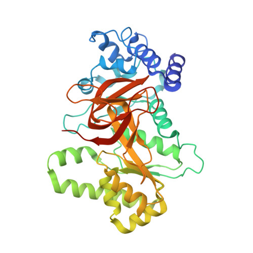
    The structure of EhpF, a 41 kDa protein that functions in the biosynthetic pathway leading to the broad-spectrum antimicrobial compound D-alanylgriseoluteic acid (AGA), is reported. A cluster of approximately 16 genes, including ehpF, located on a 200 kbp plasmid native to certain strains of Pantoea agglomerans encodes the proteins that are required for the conversion of chorismic acid to AGA. Phenazine-1,6-dicarboxylate has been identified as an intermediate in AGA biosynthesis and deletion of ehpF results in accumulation of this compound in vivo. The crystallographic data presented here reveal that EhpF is an atypical member of the acyl-CoA synthase or ANL superfamily of adenylating enzymes. These enzymes typically catalyze two-step reactions involving adenylation of a carboxylate substrate followed by transfer of the substrate from AMP to coenzyme A or another phosphopantetheine. EhpF is distinguished by the absence of the C-terminal domain that is characteristic of enzymes from this family and is involved in phosphopantetheine binding and in the second half of the canonical two-step reaction that is typically observed. Based on the structure of EhpF and a bioinformatic analysis, it is proposed that EhpF and EhpG convert phenazine-1,6-dicarboxylate to 6-formylphenazine-1-carboxylate via an adenylyl intermediate.


  • Organizational Affiliation

    Center for Advanced Research in Biotechnology, The University of Maryland Biotechnology Institute, 9600 Gudelsky Drive, Rockville, MD 20850, USA.


Macromolecules
Find similar proteins by:  (by identity cutoff)  |  3D Structure
Entity ID: 1
MoleculeChains Sequence LengthOrganismDetailsImage
EhpF
A, B
366Pantoea agglomeransMutation(s): 0 
Gene Names: ehpF
UniProt
Find proteins for Q8GPH0 (Enterobacter agglomerans)
Explore Q8GPH0 
Go to UniProtKB:  Q8GPH0
Entity Groups  
Sequence Clusters30% Identity50% Identity70% Identity90% Identity95% Identity100% Identity
UniProt GroupQ8GPH0
Sequence Annotations
Expand
  • Reference Sequence
Small Molecules
Ligands 1 Unique
IDChains Name / Formula / InChI Key2D Diagram3D Interactions
PXC
Query on PXC

Download Ideal Coordinates CCD File 
C [auth A],
D [auth B]
phenazine-1,6-dicarboxylic acid
C14 H8 N2 O4
MJALVONLCNWZHK-UHFFFAOYSA-N
Experimental Data & Validation

Experimental Data

  • Method: X-RAY DIFFRACTION
  • Resolution: 2.80 Å
  • R-Value Free: 0.288 
  • R-Value Work: 0.212 
  • R-Value Observed: 0.216 
  • Space Group: I 2 3
Unit Cell:
Length ( Å )Angle ( ˚ )
a = 193.129α = 90
b = 193.129β = 90
c = 193.129γ = 90
Software Package:
Software NamePurpose
CrystalCleardata collection
PHASERphasing
REFMACrefinement
d*TREKdata reduction
d*TREKdata scaling

Structure Validation

View Full Validation Report


Ligand Structure Quality Assessment 


Entry History 

Deposition Data

Revision History  (Full details and data files)

  • Version 1.0: 2010-05-26
    Type: Initial release
  • Version 1.1: 2011-07-13
    Changes: Advisory, Version format compliance
  • Version 1.2: 2012-03-21
    Changes: Database references
  • Version 1.3: 2023-09-06
    Changes: Data collection, Database references, Derived calculations, Refinement description