3L26

Crystal structure of Zaire Ebola VP35 interferon inhibitory domain bound to 8 bp dsRNA


Experimental Data Snapshot

  • Method: X-RAY DIFFRACTION
  • Resolution: 2.40 Å
  • R-Value Free: 0.254 
  • R-Value Work: 0.179 
  • R-Value Observed: 0.183 

wwPDB Validation   3D Report Full Report


This is version 1.2 of the entry. See complete history


Literature

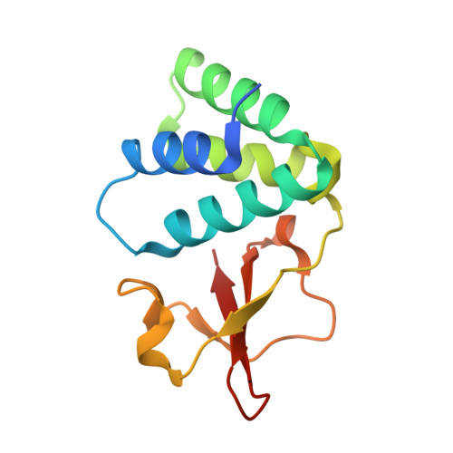
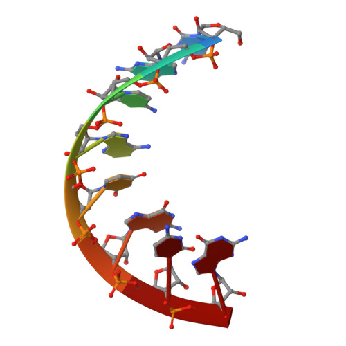
Structural basis for dsRNA recognition and interferon antagonism by Ebola VP35.

Leung, D.W.Prins, K.C.Borek, D.M.Farahbakhsh, M.Tufariello, J.M.Ramanan, P.Nix, J.C.Helgeson, L.A.Otwinowski, Z.Honzatko, R.B.Basler, C.F.Amarasinghe, G.K.

(2010) Nat Struct Mol Biol 17: 165-172

  • DOI: https://doi.org/10.1038/nsmb.1765
  • Primary Citation of Related Structures:  
    3L25, 3L26, 3L27, 3L28

  • PubMed Abstract: 

    Ebola viral protein 35 (VP35), encoded by the highly pathogenic Ebola virus, facilitates host immune evasion by antagonizing antiviral signaling pathways, including those initiated by RIG-I-like receptors. Here we report the crystal structure of the Ebola VP35 interferon inhibitory domain (IID) bound to short double-stranded RNA (dsRNA), which together with in vivo results reveals how VP35-dsRNA interactions contribute to immune evasion. Conserved basic residues in VP35 IID recognize the dsRNA backbone, whereas the dsRNA blunt ends are 'end-capped' by a pocket of hydrophobic residues that mimic RIG-I-like receptor recognition of blunt-end dsRNA. Residues critical for RNA binding are also important for interferon inhibition in vivo but not for viral polymerase cofactor function of VP35. These results suggest that simultaneous recognition of dsRNA backbone and blunt ends provides a mechanism by which Ebola VP35 antagonizes host dsRNA sensors and immune responses.


  • Organizational Affiliation

    Department of Biochemistry, Biophysics and Molecular Biology, Iowa State University, Ames, Iowa, USA.


Macromolecules

Find similar proteins by:  (by identity cutoff)  |  3D Structure
Entity ID: 1
MoleculeChains Sequence LengthOrganismDetailsImage
Polymerase cofactor VP35
A, B
129Ebola virus - Mayinga, Zaire, 1976Mutation(s): 0 
Gene Names: VP35
UniProt
Find proteins for Q05127 (Zaire ebolavirus (strain Mayinga-76))
Explore Q05127 
Go to UniProtKB:  Q05127
Entity Groups  
Sequence Clusters30% Identity50% Identity70% Identity90% Identity95% Identity100% Identity
UniProt GroupQ05127
Sequence Annotations
Expand
  • Reference Sequence

Find similar nucleic acids by:  Sequence   |   3D Structure  

Entity ID: 2
MoleculeChains LengthOrganismImage
RNA (5'-R(*CP*GP*CP*AP*UP*GP*CP*G)-3')8N/A
Sequence Annotations
Expand
  • Reference Sequence
Small Molecules
Ligands 2 Unique
IDChains Name / Formula / InChI Key2D Diagram3D Interactions
CL
Query on CL

Download Ideal Coordinates CCD File 
F [auth A],
G [auth A],
H [auth A],
K [auth B],
L [auth B]
CHLORIDE ION
Cl
VEXZGXHMUGYJMC-UHFFFAOYSA-M
MG
Query on MG

Download Ideal Coordinates CCD File 
D [auth A],
E [auth A],
I [auth B],
J [auth B],
M [auth C]
MAGNESIUM ION
Mg
JLVVSXFLKOJNIY-UHFFFAOYSA-N
Experimental Data & Validation

Experimental Data

  • Method: X-RAY DIFFRACTION
  • Resolution: 2.40 Å
  • R-Value Free: 0.254 
  • R-Value Work: 0.179 
  • R-Value Observed: 0.183 
  • Space Group: P 43 21 2
Unit Cell:
Length ( Å )Angle ( ˚ )
a = 81.025α = 90
b = 81.025β = 90
c = 90.249γ = 90
Software Package:
Software NamePurpose
CrystalCleardata collection
PHASERphasing
REFMACrefinement
d*TREKdata reduction
d*TREKdata scaling

Structure Validation

View Full Validation Report


Entry History 

Revision History  (Full details and data files)

  • Version 1.0: 2010-01-26
    Type: Initial release
  • Version 1.1: 2011-07-13
    Changes: Advisory, Version format compliance
  • Version 1.2: 2023-09-06
    Changes: Data collection, Database references, Derived calculations, Refinement description