3KJD

Human poly(ADP-ribose) polymerase 2, catalytic fragment in complex with an inhibitor ABT-888


Experimental Data Snapshot

  • Method: X-RAY DIFFRACTION
  • Resolution: 1.95 Å
  • R-Value Free: 0.232 
  • R-Value Work: 0.179 
  • R-Value Observed: 0.182 

wwPDB Validation   3D Report Full Report


Ligand Structure Quality Assessment 


This is version 1.2 of the entry. See complete history


Literature

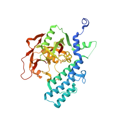
Crystal structure of the catalytic domain of human PARP2 in complex with PARP inhibitor ABT-888.

Karlberg, T.Hammarstrom, M.Schutz, P.Svensson, L.Schuler, H.

(2010) Biochemistry 49: 1056-1058

  • DOI: https://doi.org/10.1021/bi902079y
  • Primary Citation of Related Structures:  
    3KCZ, 3KJD

  • PubMed Abstract: 

    Poly-ADP-ribose polymerases (PARPs) catalyze transfer of ADP-ribose from NAD(+) to specific residues in their substrate proteins or to growing ADP-ribose chains. PARP activity is involved in processes such as chromatin remodeling, transcription control, and DNA repair. Inhibitors of PARP activity may be useful in cancer therapy. PARP2 is the family member that is most similar to PARP1, and the two can act together as heterodimers. We used X-ray crystallography to determine two structures of the catalytic domain of human PARP2: the complexes with PARP inhibitors 3-aminobenzamide and ABT-888. These results contribute to our understanding of structural features and compound properties that can be employed to develop selective inhibitors of human ADP-ribosyltransferases.


  • Organizational Affiliation

    Structural Genomics Consortium, Department of Medical Biochemistry and Biophysics, Karolinska Institutet, Scheeles vag 2, 17177 Stockholm, Sweden.


Macromolecules
Find similar proteins by:  (by identity cutoff)  |  3D Structure
Entity ID: 1
MoleculeChains Sequence LengthOrganismDetailsImage
Poly [ADP-ribose] polymerase 2
A, B
368Homo sapiensMutation(s): 0 
Gene Names: ADPRT2ADPRTL2PARP2
EC: 2.4.2.30
UniProt & NIH Common Fund Data Resources
Find proteins for Q9UGN5 (Homo sapiens)
Explore Q9UGN5 
Go to UniProtKB:  Q9UGN5
PHAROS:  Q9UGN5
GTEx:  ENSG00000129484 
Entity Groups  
Sequence Clusters30% Identity50% Identity70% Identity90% Identity95% Identity100% Identity
UniProt GroupQ9UGN5
Sequence Annotations
Expand
  • Reference Sequence
Small Molecules
Binding Affinity Annotations 
IDSourceBinding Affinity
78P BindingDB:  3KJD Kd: 5.8 (nM) from 1 assay(s)
IC50: min: 1.3, max: 138 (nM) from 8 assay(s)
Binding MOAD:  3KJD Kd: 2.9 (nM) from 1 assay(s)
PDBBind:  3KJD Kd: 2.9 (nM) from 1 assay(s)
Experimental Data & Validation

Experimental Data

  • Method: X-RAY DIFFRACTION
  • Resolution: 1.95 Å
  • R-Value Free: 0.232 
  • R-Value Work: 0.179 
  • R-Value Observed: 0.182 
  • Space Group: P 1 21 1
Unit Cell:
Length ( Å )Angle ( ˚ )
a = 58.134α = 90
b = 134.611β = 117.68
c = 58.314γ = 90
Software Package:
Software NamePurpose
DNAdata collection
MOLREPphasing
REFMACrefinement
XDSdata reduction
XSCALEdata scaling

Structure Validation

View Full Validation Report


Ligand Structure Quality Assessment 


Entry History 

Revision History  (Full details and data files)

  • Version 1.0: 2009-11-17
    Type: Initial release
  • Version 1.1: 2011-07-13
    Changes: Non-polymer description, Version format compliance
  • Version 1.2: 2023-11-01
    Changes: Data collection, Database references, Derived calculations, Refinement description, Structure summary