3KIO

mouse RNase H2 complex


Experimental Data Snapshot

  • Method: X-RAY DIFFRACTION
  • Resolution: 2.90 Å
  • R-Value Free: 0.305 
  • R-Value Work: 0.234 
  • R-Value Observed: 0.238 

wwPDB Validation   3D Report Full Report


This is version 1.1 of the entry. See complete history

Re-refinement Note

A newer entry is available that reflects an alternative modeling of the original data: 3P5J


Literature

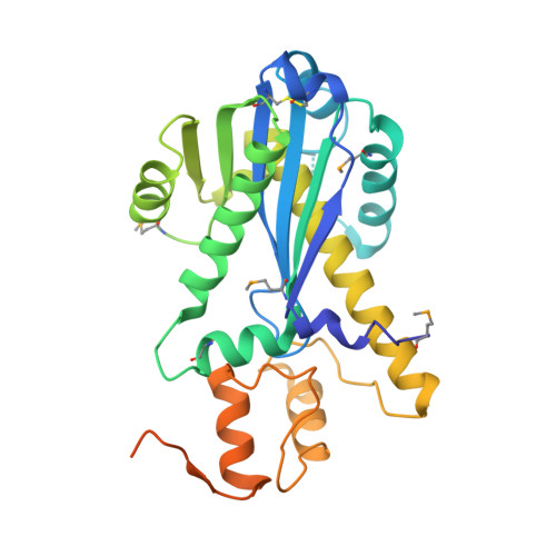
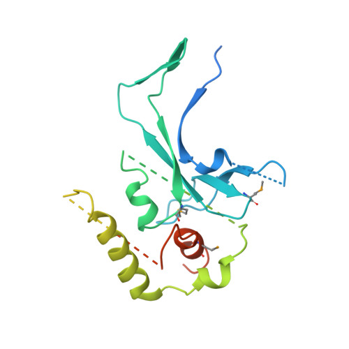
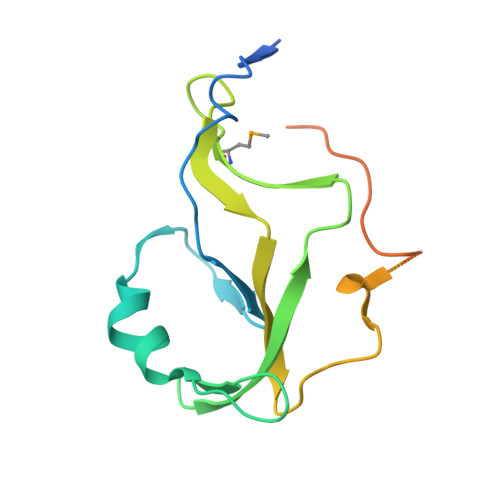
The structure of the mammalian RNase H2 complex provides insight into RNA:DNA hybrid processing to prevent immune dysfunction.

Shaban, N.M.Harvey, S.Perrino, F.W.Hollis, T.

(2010) J Biol Chem 285: 3617-3624

  • DOI: https://doi.org/10.1074/jbc.M109.059048
  • Primary Citation of Related Structures:  
    3KIO

  • PubMed Abstract: 

    The mammalian RNase H2 ribonuclease complex has a critical function in nucleic acid metabolism to prevent immune activation with likely roles in processing of RNA primers in Okazaki fragments during DNA replication, in removing ribonucleotides misinserted by DNA polymerases, and in eliminating RNA.DNA hybrids during cell death. Mammalian RNase H2 is a heterotrimeric complex of the RNase H2A, RNase H2B, and RNase H2C proteins that are all required for proper function and activity. Mutations in the human RNase H2 genes cause Aicardi-Goutières syndrome. We have determined the crystal structure of the three-protein mouse RNase H2 enzyme complex to better understand the molecular basis of RNase H2 dysfunction in human autoimmunity. The structure reveals the intimately interwoven architecture of RNase H2B and RNase H2C that interface with RNase H2A in a complex ideally suited for nucleic acid binding and hydrolysis coupled to protein-protein interaction motifs that could allow for efficient participation in multiple cellular functions. We have identified four conserved acidic residues in the active site that are necessary for activity and suggest a two-metal ion mechanism of catalysis for RNase H2. An Okazaki fragment has been modeled into the RNase H2 nucleic acid binding site providing insight into the recognition of RNA.DNA junctions by the RNase H2. Further structural and biochemical analyses show that some RNase H2 disease-causing mutations likely result in aberrant protein-protein interactions while the RNase H2A subunit-G37S mutation appears to distort the active site accounting for the demonstrated substrate specificity modification.


  • Organizational Affiliation

    From the Department of Biochemistry, Center for Structural Biology, Wake Forest University School of Medicine, Winston-Salem, North Carolina 27157.


Macromolecules
Find similar proteins by:  (by identity cutoff)  |  3D Structure
Entity ID: 1
MoleculeChains Sequence LengthOrganismDetailsImage
Ribonuclease H2 subunit A301Mus musculusMutation(s): 0 
Gene Names: RNase H2ARnaseh2aRnasehi
EC: 3.1.26.4
UniProt & NIH Common Fund Data Resources
Find proteins for Q9CWY8 (Mus musculus)
Explore Q9CWY8 
Go to UniProtKB:  Q9CWY8
IMPC:  MGI:1916974
Entity Groups  
Sequence Clusters30% Identity50% Identity70% Identity90% Identity95% Identity100% Identity
UniProt GroupQ9CWY8
Sequence Annotations
Expand
  • Reference Sequence
Find similar proteins by:  (by identity cutoff)  |  3D Structure
Entity ID: 2
MoleculeChains Sequence LengthOrganismDetailsImage
Ribonuclease H2 subunit B332Mus musculusMutation(s): 1 
Gene Names: Dleu8RNase H2BRnaseh2b
UniProt
Find proteins for Q80ZV0 (Mus musculus)
Explore Q80ZV0 
Go to UniProtKB:  Q80ZV0
Entity Groups  
Sequence Clusters30% Identity50% Identity70% Identity90% Identity95% Identity100% Identity
UniProt GroupQ80ZV0
Sequence Annotations
Expand
  • Reference Sequence
Find similar proteins by:  (by identity cutoff)  |  3D Structure
Entity ID: 3
MoleculeChains Sequence LengthOrganismDetailsImage
Ribonuclease H2 subunit C166Mus musculusMutation(s): 1 
Gene Names: Ayp1RNase H2CRnaseh2c
UniProt & NIH Common Fund Data Resources
Find proteins for Q9CQ18 (Mus musculus)
Explore Q9CQ18 
Go to UniProtKB:  Q9CQ18
IMPC:  MGI:1915459
Entity Groups  
Sequence Clusters30% Identity50% Identity70% Identity90% Identity95% Identity100% Identity
UniProt GroupQ9CQ18
Sequence Annotations
Expand
  • Reference Sequence
Small Molecules
Modified Residues  1 Unique
IDChains TypeFormula2D DiagramParent
MSE
Query on MSE
A
L-PEPTIDE LINKINGC5 H11 N O2 SeMET
Experimental Data & Validation

Experimental Data

  • Method: X-RAY DIFFRACTION
  • Resolution: 2.90 Å
  • R-Value Free: 0.305 
  • R-Value Work: 0.234 
  • R-Value Observed: 0.238 
  • Space Group: P 21 21 2
Unit Cell:
Length ( Å )Angle ( ˚ )
a = 279.287α = 90
b = 40.421β = 90
c = 67.82γ = 90
Software Package:
Software NamePurpose
ADSCdata collection
MLPHAREphasing
PHENIXrefinement
HKL-2000data reduction
HKL-2000data scaling

Structure Validation

View Full Validation Report


Entry History 

Deposition Data

Revision History  (Full details and data files)

  • Version 1.0: 2009-11-17
    Type: Initial release
  • Version 1.1: 2011-07-13
    Changes: Version format compliance