3KH2

Crystal structure of the P1 bacteriophage Doc toxin (F68S) in complex with the Phd antitoxin (L17M/V39A). Northeast Structural Genomics targets ER385-ER386


Experimental Data Snapshot

  • Method: X-RAY DIFFRACTION
  • Resolution: 2.71 Å
  • R-Value Free: 0.267 
  • R-Value Work: 0.217 
  • R-Value Observed: 0.219 

wwPDB Validation   3D Report Full Report


Ligand Structure Quality Assessment 


This is version 1.5 of the entry. See complete history


Literature

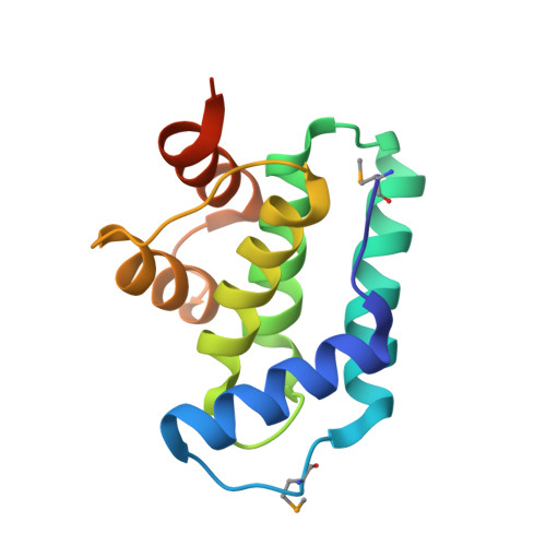
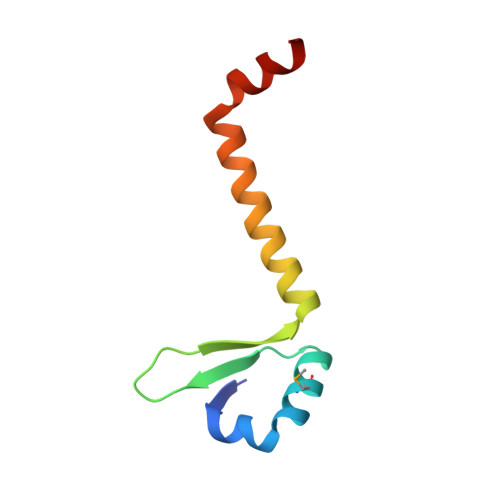
Crystal Structures of Phd-Doc, HigA, and YeeU Establish Multiple Evolutionary Links between Microbial Growth-Regulating Toxin-Antitoxin Systems.

Arbing, M.A.Handelman, S.K.Kuzin, A.P.Verdon, G.Wang, C.Su, M.Rothenbacher, F.P.Abashidze, M.Liu, M.Hurley, J.M.Xiao, R.Acton, T.Inouye, M.Montelione, G.T.Woychik, N.A.Hunt, J.F.

(2010) Structure 18: 996-1010

  • DOI: https://doi.org/10.1016/j.str.2010.04.018
  • Primary Citation of Related Structures:  
    2H28, 2ICT, 2INW, 3KH2

  • PubMed Abstract: 

    Bacterial toxin-antitoxin (TA) systems serve a variety of physiological functions including regulation of cell growth and maintenance of foreign genetic elements. Sequence analyses suggest that TA families are linked by complex evolutionary relationships reflecting likely swapping of functional domains between different TA families. Our crystal structures of Phd-Doc from bacteriophage P1, the HigA antitoxin from Escherichia coli CFT073, and YeeU of the YeeUWV systems from E. coli K12 and Shigella flexneri confirm this inference and reveal additional, unanticipated structural relationships. The growth-regulating Doc toxin exhibits structural similarity to secreted virulence factors that are toxic for eukaryotic target cells. The Phd antitoxin possesses the same fold as both the YefM and NE2111 antitoxins that inhibit structurally unrelated toxins. YeeU, which has an antitoxin-like activity that represses toxin expression, is structurally similar to the ribosome-interacting toxins YoeB and RelE. These observations suggest extensive functional exchanges have occurred between TA systems during bacterial evolution.


  • Organizational Affiliation

    Department of Biological Sciences, Columbia University, 702 Fairchild Center, MC2434, New York, NY 10027, USA.


Macromolecules
Find similar proteins by:  (by identity cutoff)  |  3D Structure
Entity ID: 1
MoleculeChains Sequence LengthOrganismDetailsImage
Death on curing protein
A, B, C, D
134Punavirus P1Mutation(s): 1 
Gene Names: doc
UniProt
Find proteins for Q06259 (Escherichia phage P1)
Explore Q06259 
Go to UniProtKB:  Q06259
Entity Groups  
Sequence Clusters30% Identity50% Identity70% Identity90% Identity95% Identity100% Identity
UniProt GroupQ06259
Sequence Annotations
Expand
  • Reference Sequence
Find similar proteins by:  (by identity cutoff)  |  3D Structure
Entity ID: 2
MoleculeChains Sequence LengthOrganismDetailsImage
Prevent host death protein
E, F, G, H
73Punavirus P1Mutation(s): 2 
Gene Names: phd
UniProt
Find proteins for Q06253 (Escherichia phage P1)
Explore Q06253 
Go to UniProtKB:  Q06253
Entity Groups  
Sequence Clusters30% Identity50% Identity70% Identity90% Identity95% Identity100% Identity
UniProt GroupQ06253
Sequence Annotations
Expand
  • Reference Sequence
Small Molecules
Ligands 4 Unique
IDChains Name / Formula / InChI Key2D Diagram3D Interactions
HED
Query on HED

Download Ideal Coordinates CCD File 
J [auth A],
M [auth B]
2-HYDROXYETHYL DISULFIDE
C4 H10 O2 S2
KYNFOMQIXZUKRK-UHFFFAOYSA-N
PO4
Query on PO4

Download Ideal Coordinates CCD File 
N [auth E],
O [auth F],
P [auth G],
R [auth H]
PHOSPHATE ION
O4 P
NBIIXXVUZAFLBC-UHFFFAOYSA-K
GOL
Query on GOL

Download Ideal Coordinates CCD File 
K [auth B],
L [auth B],
Q [auth H]
GLYCEROL
C3 H8 O3
PEDCQBHIVMGVHV-UHFFFAOYSA-N
CL
Query on CL

Download Ideal Coordinates CCD File 
I [auth A]CHLORIDE ION
Cl
VEXZGXHMUGYJMC-UHFFFAOYSA-M
Modified Residues  1 Unique
IDChains TypeFormula2D DiagramParent
MSE
Query on MSE
A, B, C, D
L-PEPTIDE LINKINGC5 H11 N O2 SeMET
Experimental Data & Validation

Experimental Data

  • Method: X-RAY DIFFRACTION
  • Resolution: 2.71 Å
  • R-Value Free: 0.267 
  • R-Value Work: 0.217 
  • R-Value Observed: 0.219 
  • Space Group: P 21 21 21
Unit Cell:
Length ( Å )Angle ( ˚ )
a = 95.899α = 90
b = 111.277β = 90
c = 118.747γ = 90
Software Package:
Software NamePurpose
DENZOdata reduction
SCALEPACKdata scaling
COMOphasing
REFMACrefinement
PDB_EXTRACTdata extraction

Structure Validation

View Full Validation Report


Ligand Structure Quality Assessment 


Entry History 

Revision History  (Full details and data files)

  • Version 1.0: 2010-08-18
    Type: Initial release
  • Version 1.1: 2011-07-13
    Changes: Version format compliance
  • Version 1.2: 2017-11-01
    Changes: Advisory, Refinement description
  • Version 1.3: 2021-10-13
    Changes: Advisory, Database references, Derived calculations
  • Version 1.4: 2023-09-06
    Changes: Data collection, Refinement description
  • Version 1.5: 2023-11-22
    Changes: Data collection