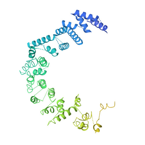
Crystal structure of DNA-PKcs reveals a large open-ring cradle comprised of HEAT repeats.
Sibanda, B.L., Chirgadze, D.Y., Blundell, T.L.(2010) Nature 463: 118-121
- PubMed: 20023628
- DOI: https://doi.org/10.1038/nature08648
- Primary Citation of Related Structures:
3KGV - PubMed Abstract:
Broken chromosomes arising from DNA double-strand breaks result from endogenous events such as the production of reactive oxygen species during cellular metabolism, as well as from exogenous sources such as ionizing radiation. Left unrepaired or incorrectly repaired they can lead to genomic changes that may result in cell death or cancer. DNA-dependent protein kinase (DNA-PK), a holoenzyme that comprises the DNA-PK catalytic subunit (DNA-PKcs) and the heterodimer Ku70/Ku80, has a major role in non-homologous end joining-the main pathway in mammals used to repair double-strand breaks. DNA-PKcs is a serine/threonine protein kinase comprising a single polypeptide chain of 4,128 amino acids and belonging to the phosphatidylinositol-3-OH kinase (PI(3)K)-related protein family. DNA-PKcs is involved in the sensing and transmission of DNA damage signals to proteins such as p53, setting off events that lead to cell cycle arrest. It phosphorylates a wide range of substrates in vitro, including Ku70/Ku80, which is translocated along DNA. Here we present the crystal structure of human DNA-PKcs at 6.6 A resolution, in which the overall fold is clearly visible, to our knowledge, for the first time. The many alpha-helical HEAT repeats (helix-turn-helix motifs) facilitate bending and allow the polypeptide chain to fold into a hollow circular structure. The carboxy-terminal kinase domain is located on top of this structure, and a small HEAT repeat domain that probably binds DNA is inside. The structure provides a flexible cradle to promote DNA double-strand-break repair.
Organizational Affiliation:
Department of Biochemistry, University of Cambridge, Old Addenbrooke's site, 80 Tennis Court Road, Cambridge CB2 1GA, UK. lynn@cryst.bioc.cam.ac.uk