3K4C

Pyranose 2-oxidase H167A/T169G mutant


Experimental Data Snapshot

  • Method: X-RAY DIFFRACTION
  • Resolution: 1.70 Å
  • R-Value Free: 0.216 
  • R-Value Work: 0.180 
  • R-Value Observed: 0.181 

wwPDB Validation   3D Report Full Report


Ligand Structure Quality Assessment 


This is version 1.5 of the entry. See complete history


Literature

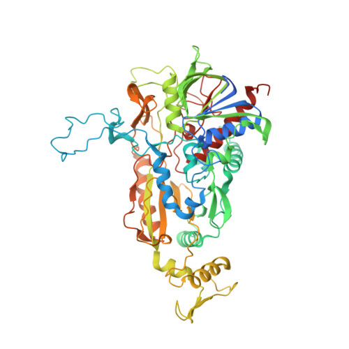
A conserved active-site threonine is important for both sugar and flavin oxidations of pyranose 2-oxidase.

Pitsawong, W.Sucharitakul, J.Prongjit, M.Tan, T.C.Spadiut, O.Haltrich, D.Divne, C.Chaiyen, P.

(2010) J Biol Chem 285: 9697-9705

  • DOI: https://doi.org/10.1074/jbc.M109.073247
  • Primary Citation of Related Structures:  
    3K4B, 3K4C

  • PubMed Abstract: 

    Pyranose 2-oxidase (P2O) catalyzes the oxidation by O(2) of d-glucose and several aldopyranoses to yield the 2-ketoaldoses and H(2)O(2). Based on crystal structures, in one rotamer conformation, the threonine hydroxyl of Thr(169) forms H-bonds to the flavin-N5/O4 locus, whereas, in a different rotamer, it may interact with either sugar or other parts of the P2O.sugar complex. Transient kinetics of wild-type (WT) and Thr(169) --> S/N/G/A replacement variants show that D-Glc binds to T169S, T169N, and WT with the same K(d) (45-47 mm), and the hydride transfer rate constants (k(red)) are similar (15.3-9.7 s(-1) at 4 degrees C). k(red) of T169G with D-glucose (0.7 s(-1), 4 degrees C) is significantly less than that of WT but not as severely affected as in T169A (k(red) of 0.03 s(-1) at 25 degrees C). Transient kinetics of WT and mutants using d-galactose show that P2O binds d-galactose with a one-step binding process, different from binding of d-glucose. In T169S, T169N, and T169G, the overall turnover with d-Gal is faster than that of WT due to an increase of k(red). In the crystal structure of T169S, Ser(169) O gamma assumes a position identical to that of O gamma 1 in Thr(169); in T169G, solvent molecules may be able to rescue H-bonding. Our data suggest that a competent reductive half-reaction requires a side chain at position 169 that is able to form an H-bond within the ES complex. During the oxidative half-reaction, all mutants failed to stabilize a C4a-hydroperoxyflavin intermediate, thus suggesting that the precise position and geometry of the Thr(169) side chain are required for intermediate stabilization.


  • Organizational Affiliation

    Department of Biochemistry and Center of Excellence in Protein Structure and Function, Faculty of Science, Mahidol University, Bangkok 10400, Thailand.


Macromolecules
Find similar proteins by:  (by identity cutoff)  |  3D Structure
Entity ID: 1
MoleculeChains Sequence LengthOrganismDetailsImage
Pyranose 2-oxidase
A, B, C, D
623Trametes ochraceaMutation(s): 2 
Gene Names: p2o
EC: 1.1.3.10
UniProt
Find proteins for Q7ZA32 (Trametes ochracea)
Explore Q7ZA32 
Go to UniProtKB:  Q7ZA32
Entity Groups  
Sequence Clusters30% Identity50% Identity70% Identity90% Identity95% Identity100% Identity
UniProt GroupQ7ZA32
Sequence Annotations
Expand
  • Reference Sequence
Small Molecules
Ligands 3 Unique
IDChains Name / Formula / InChI Key2D Diagram3D Interactions
FAD
Query on FAD

Download Ideal Coordinates CCD File 
E [auth A],
H [auth B],
K [auth C],
M [auth D]
FLAVIN-ADENINE DINUCLEOTIDE
C27 H33 N9 O15 P2
VWWQXMAJTJZDQX-UYBVJOGSSA-N
1PE
Query on 1PE

Download Ideal Coordinates CCD File 
F [auth A],
G [auth A],
J [auth B],
N [auth D]
PENTAETHYLENE GLYCOL
C10 H22 O6
JLFNLZLINWHATN-UHFFFAOYSA-N
MES
Query on MES

Download Ideal Coordinates CCD File 
I [auth B],
L [auth C]
2-(N-MORPHOLINO)-ETHANESULFONIC ACID
C6 H13 N O4 S
SXGZJKUKBWWHRA-UHFFFAOYSA-N
Experimental Data & Validation

Experimental Data

  • Method: X-RAY DIFFRACTION
  • Resolution: 1.70 Å
  • R-Value Free: 0.216 
  • R-Value Work: 0.180 
  • R-Value Observed: 0.181 
  • Space Group: P 1 21 1
Unit Cell:
Length ( Å )Angle ( ˚ )
a = 100.206α = 90
b = 102.964β = 90.82
c = 137.122γ = 90
Software Package:
Software NamePurpose
MAR345data collection
REFMACrefinement
XDSdata reduction
XSCALEdata scaling
REFMACphasing

Structure Validation

View Full Validation Report


Ligand Structure Quality Assessment 


Entry History 

Deposition Data

Revision History  (Full details and data files)

  • Version 1.0: 2010-02-02
    Type: Initial release
  • Version 1.1: 2011-07-13
    Changes: Advisory, Version format compliance
  • Version 1.2: 2014-11-12
    Changes: Structure summary
  • Version 1.3: 2017-11-01
    Changes: Data collection, Refinement description
  • Version 1.4: 2021-10-13
    Changes: Database references, Derived calculations
  • Version 1.5: 2023-09-06
    Changes: Data collection, Refinement description