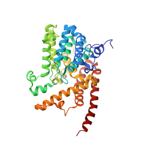
3JSW

Human PDE9 in complex with selective inhibitor


Experimental Data Snapshot

  • Method: X-RAY DIFFRACTION
  • Resolution: 2.30 Å
  • R-Value Free: 0.212 
  • R-Value Work: 0.179 
  • R-Value Observed: 0.183 

wwPDB Validation   3D Report Full Report


Ligand Structure Quality Assessment 


This is version 1.3 of the entry. See complete history


Literature

Identification of a Brain Penetrant PDE9A Inhibitor Utilizing Prospective Design and Chemical Enablement as a Rapid Lead Optimization Strategy.

Verhoest, P.R.Proulx-Lafrance, C.Corman, M.Chenard, L.Helal, C.J.Hou, X.Kleiman, R.Liu, S.Marr, E.Menniti, F.S.Schmidt, C.J.Vanase-Frawley, M.Schmidt, A.W.Williams, R.D.Nelson, F.R.Fonseca, K.R.Liras, S.

(2009) J Med Chem 52: 7946-7949

  • DOI: https://doi.org/10.1021/jm9015334
  • Primary Citation of Related Structures:  
    3JSI, 3JSW

  • PubMed Abstract: 

    By use of chemical enablement and prospective design, a novel series of selective, brain penetrant PDE9A inhibitors have been identified that are capable of producing in vivo elevations of brain cGMP.


  • Organizational Affiliation

    Neuroscience Chemistry, Pfizer Global Research and Development, Groton, CT 06340, USA.


Macromolecules
Find similar proteins by:  (by identity cutoff)  |  3D Structure
Entity ID: 1
MoleculeChains Sequence LengthOrganismDetailsImage
High affinity cGMP-specific 3',5'-cyclic phosphodiesterase 9A
A, B
329Homo sapiensMutation(s): 0 
Gene Names: PDE9A
EC: 3.1.4.35
UniProt & NIH Common Fund Data Resources
Find proteins for O76083 (Homo sapiens)
Explore O76083 
Go to UniProtKB:  O76083
PHAROS:  O76083
GTEx:  ENSG00000160191 
Entity Groups  
Sequence Clusters30% Identity50% Identity70% Identity90% Identity95% Identity100% Identity
UniProt GroupO76083
Sequence Annotations
Expand
  • Reference Sequence
Small Molecules
Binding Affinity Annotations 
IDSourceBinding Affinity
JAR PDBBind:  3JSW IC50: 66 (nM) from 1 assay(s)
Binding MOAD:  3JSW IC50: 66 (nM) from 1 assay(s)
BindingDB:  3JSW IC50: 66 (nM) from 1 assay(s)
Experimental Data & Validation

Experimental Data

  • Method: X-RAY DIFFRACTION
  • Resolution: 2.30 Å
  • R-Value Free: 0.212 
  • R-Value Work: 0.179 
  • R-Value Observed: 0.183 
  • Space Group: P 41 21 2
Unit Cell:
Length ( Å )Angle ( ˚ )
a = 103.278α = 90
b = 103.278β = 90
c = 270.349γ = 90
Software Package:
Software NamePurpose
REFMACrefinement
HKL-2000data reduction
HKL-2000data scaling
REFMACphasing

Structure Validation

View Full Validation Report


Ligand Structure Quality Assessment 


Entry History 

Deposition Data

  • Released Date: 2009-12-01 
  • Deposition Author(s): Liu, S.

Revision History  (Full details and data files)

  • Version 1.0: 2009-12-01
    Type: Initial release
  • Version 1.1: 2011-07-13
    Changes: Advisory, Version format compliance
  • Version 1.2: 2017-11-01
    Changes: Refinement description
  • Version 1.3: 2023-09-06
    Changes: Data collection, Database references, Derived calculations, Refinement description