3JQO

Crystal structure of the outer membrane complex of a type IV secretion system


Experimental Data Snapshot

  • Method: X-RAY DIFFRACTION
  • Resolution: 2.60 Å
  • R-Value Free: 0.259 
  • R-Value Work: 0.227 
  • R-Value Observed: 0.229 

wwPDB Validation   3D Report Full Report


Ligand Structure Quality Assessment 


This is version 1.5 of the entry. See complete history


Literature

Structure of the outer membrane complex of a type IV secretion system

Chandran, V.Fronzes, R.Duquerroy, S.Cronin, N.Navaza, J.Waksman, G.

(2009) Nature 462: 1011-1015

  • DOI: https://doi.org/10.1038/nature08588
  • Primary Citation of Related Structures:  
    3JQO

  • PubMed Abstract: 

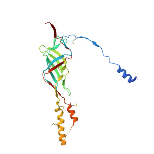
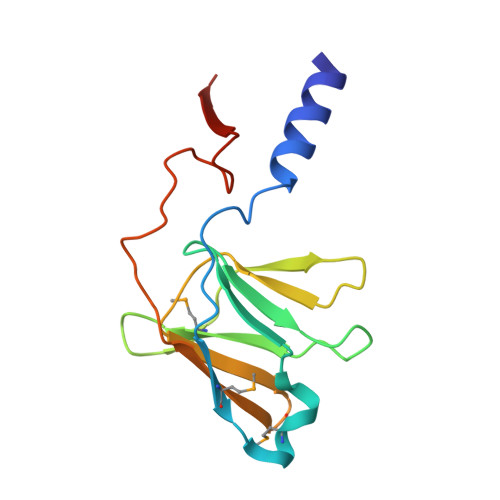
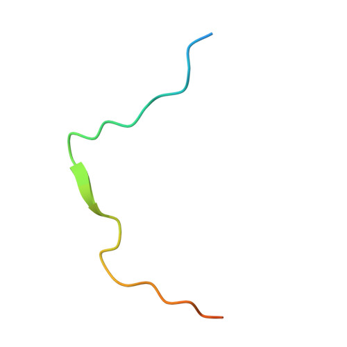
    Type IV secretion systems are secretion nanomachines spanning the two membranes of Gram-negative bacteria. Three proteins, VirB7, VirB9 and VirB10, assemble into a 1.05 megadalton (MDa) core spanning the inner and outer membranes. This core consists of 14 copies of each of the proteins and forms two layers, the I and O layers, inserting in the inner and outer membrane, respectively. Here we present the crystal structure of a approximately 0.6 MDa outer-membrane complex containing the entire O layer. This structure is the largest determined for an outer-membrane channel and is unprecedented in being composed of three proteins. Unexpectedly, this structure identifies VirB10 as the outer-membrane channel with a unique hydrophobic double-helical transmembrane region. This structure establishes VirB10 as the only known protein crossing both membranes of Gram-negative bacteria. Comparison of the cryo-electron microscopy (cryo-EM) and crystallographic structures points to conformational changes regulating channel opening and closing.


  • Organizational Affiliation

    Institute of Structural and Molecular Biology, University College London and Birkbeck College, Malet Street, London WC1E 7HX, UK.


Macromolecules
Find similar proteins by:  (by identity cutoff)  |  3D Structure
Entity ID: 1
MoleculeChains Sequence LengthOrganismDetailsImage
TraF protein227Escherichia coli K-12Mutation(s): 0 
Gene Names: traF
Membrane Entity: Yes 
UniProt
Find proteins for Q46705 (Escherichia coli)
Explore Q46705 
Go to UniProtKB:  Q46705
Entity Groups  
Sequence Clusters30% Identity50% Identity70% Identity90% Identity95% Identity100% Identity
UniProt GroupQ46705
Sequence Annotations
Expand
  • Reference Sequence
Find similar proteins by:  (by identity cutoff)  |  3D Structure
Entity ID: 2
MoleculeChains Sequence LengthOrganismDetailsImage
TraO protein135Escherichia coli K-12Mutation(s): 0 
Gene Names: traO
Membrane Entity: Yes 
UniProt
Find proteins for Q46704 (Escherichia coli)
Explore Q46704 
Go to UniProtKB:  Q46704
Entity Groups  
Sequence Clusters30% Identity50% Identity70% Identity90% Identity95% Identity100% Identity
UniProt GroupQ46704
Sequence Annotations
Expand
  • Reference Sequence
Find similar proteins by:  (by identity cutoff)  |  3D Structure
Entity ID: 3
MoleculeChains Sequence LengthOrganismDetailsImage
TraN protein34Escherichia coli K-12Mutation(s): 0 
Gene Names: traN
UniProt
Find proteins for Q46702 (Escherichia coli)
Explore Q46702 
Go to UniProtKB:  Q46702
Entity Groups  
Sequence Clusters30% Identity50% Identity70% Identity90% Identity95% Identity100% Identity
UniProt GroupQ46702
Sequence Annotations
Expand
  • Reference Sequence
Small Molecules
Modified Residues  1 Unique
IDChains TypeFormula2D DiagramParent
MSE
Query on MSE
A
BA [auth b]
D
EA [auth e]
G
A,
BA [auth b],
D,
EA [auth e],
G,
HA [auth h],
J,
KA [auth k],
M,
NA [auth n],
P,
S,
V,
Y
L-PEPTIDE LINKINGC5 H11 N O2 SeMET
Experimental Data & Validation

Experimental Data

  • Method: X-RAY DIFFRACTION
  • Resolution: 2.60 Å
  • R-Value Free: 0.259 
  • R-Value Work: 0.227 
  • R-Value Observed: 0.229 
  • Space Group: P 21 21 2
Unit Cell:
Length ( Å )Angle ( ˚ )
a = 202.4α = 90
b = 211.63β = 90
c = 203.44γ = 90
Software Package:
Software NamePurpose
ADSCdata collection
AMoREphasing
REFMACrefinement
XDSdata reduction
XSCALEdata scaling

Structure Validation

View Full Validation Report


Ligand Structure Quality Assessment 


Entry History 

Deposition Data

Revision History  (Full details and data files)

  • Version 1.0: 2009-12-01
    Type: Initial release
  • Version 1.1: 2011-07-13
    Changes: Version format compliance
  • Version 1.2: 2014-02-12
    Changes: Database references
  • Version 1.3: 2014-07-23
    Changes: Other
  • Version 1.4: 2019-11-20
    Changes: Database references, Derived calculations
  • Version 1.5: 2024-04-03
    Changes: Data collection, Database references, Derived calculations, Refinement description