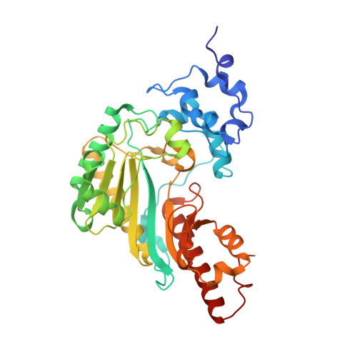
The structure of lombricine kinase: implications for phosphagen kinase conformational changes.
Bush, D.J., Kirillova, O., Clark, S.A., Davulcu, O., Fabiola, F., Xie, Q., Somasundaram, T., Ellington, W.R., Chapman, M.S.(2011) J Biol Chem 286: 9338-9350
- PubMed: 21212263
- DOI: https://doi.org/10.1074/jbc.M110.202796
- Primary Citation of Related Structures:
3JPZ, 3JQ3 - PubMed Abstract:
Lombricine kinase is a member of the phosphagen kinase family and a homolog of creatine and arginine kinases, enzymes responsible for buffering cellular ATP levels. Structures of lombricine kinase from the marine worm Urechis caupo were determined by x-ray crystallography. One form was crystallized as a nucleotide complex, and the other was substrate-free. The two structures are similar to each other and more similar to the substrate-free forms of homologs than to the substrate-bound forms of the other phosphagen kinases. Active site specificity loop 309-317, which is disordered in substrate-free structures of homologs and is known from the NMR of arginine kinase to be inherently dynamic, is resolved in both lombricine kinase structures, providing an improved basis for understanding the loop dynamics. Phosphagen kinases undergo a segmented closing on substrate binding, but the lombricine kinase ADP complex is in the open form more typical of substrate-free homologs. Through a comparison with prior complexes of intermediate structure, a correlation was revealed between the overall enzyme conformation and the substrate interactions of His(178). Comparative modeling provides a rationale for the more relaxed specificity of these kinases, of which the natural substrates are among the largest of the phosphagen substrates.
Organizational Affiliation:
Department of Chemistry and Biochemistry, Florida State University, Tallahassee, Florida 32306, USA.