3J91

Cryo-electron microscopy of Enterovirus 71 (EV71) procapsid in complex with Fab fragments of neutralizing antibody 22A12


Experimental Data Snapshot

  • Method: ELECTRON MICROSCOPY
  • Resolution: 8.80 Å
  • Aggregation State: PARTICLE 
  • Reconstruction Method: SINGLE PARTICLE 

wwPDB Validation   3D Report Full Report


This is version 1.3 of the entry. See complete history


Literature

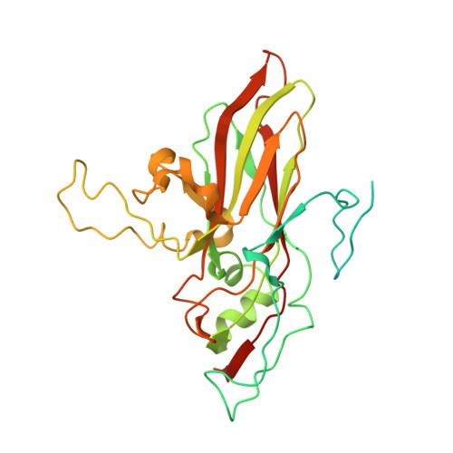
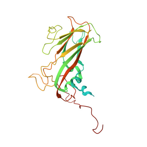
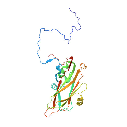
The enterovirus 71 procapsid binds neutralizing antibodies and rescues virus infection in vitro.

Shingler, K.L.Cifuente, J.O.Ashley, R.E.Makhov, A.M.Conway, J.F.Hafenstein, S.

(2015) J Virol 89: 1900-1908

  • DOI: https://doi.org/10.1128/JVI.03098-14
  • Primary Citation of Related Structures:  
    3J91, 3J93

  • PubMed Abstract: 

    Enterovirus 71 (EV71) is responsible for seasonal outbreaks of hand, foot, and mouth disease in the Asia-Pacific region. The virus has the capability to cause severe disease and death, especially in young children. Although several vaccines are currently in clinical trials, no vaccines or therapeutics have been approved for use. Previous structural studies have revealed that two antigenically distinct capsid forms are produced in EV71-infected cells: an expanded empty capsid, sometimes called a procapsid, and the infectious virus. Specifically, an immunodominant epitope of EV71 that maps to the virus canyon is structurally different in the procapsid and virus. This structure-function study shows that the procapsid can sequester antibodies, thus enhancing EV71 infection in vitro. The results presented here suggest that, due to conformational differences between the EV71 procapsid and virus, the presence of the procapsid in natural virus infections should be considered in the future design of vaccines or therapeutics.


  • Organizational Affiliation

    Department of Medicine and Department of Microbiology and Immunology, The Pennsylvania State University College of Medicine, Hershey, Pennsylvania, USA.


Macromolecules
Find similar proteins by:  (by identity cutoff)  |  3D Structure
Entity ID: 1
MoleculeChains Sequence LengthOrganismDetailsImage
VP0A [auth 0]323Enterovirus A71Mutation(s): 0 
UniProt
Find proteins for E5RPG0 (Human enterovirus 71)
Explore E5RPG0 
Go to UniProtKB:  E5RPG0
Entity Groups  
Sequence Clusters30% Identity50% Identity70% Identity90% Identity95% Identity100% Identity
UniProt GroupE5RPG0
Sequence Annotations
Expand
  • Reference Sequence
Find similar proteins by:  (by identity cutoff)  |  3D Structure
Entity ID: 2
MoleculeChains Sequence LengthOrganismDetailsImage
VP1B [auth 1]297Enterovirus A71Mutation(s): 0 
UniProt
Find proteins for E5RPG0 (Human enterovirus 71)
Explore E5RPG0 
Go to UniProtKB:  E5RPG0
Entity Groups  
Sequence Clusters30% Identity50% Identity70% Identity90% Identity95% Identity100% Identity
UniProt GroupE5RPG0
Sequence Annotations
Expand
  • Reference Sequence
Find similar proteins by:  (by identity cutoff)  |  3D Structure
Entity ID: 3
MoleculeChains Sequence LengthOrganismDetailsImage
VP3C [auth 3]242Enterovirus A71Mutation(s): 0 
UniProt
Find proteins for E5RPG0 (Human enterovirus 71)
Explore E5RPG0 
Go to UniProtKB:  E5RPG0
Entity Groups  
Sequence Clusters30% Identity50% Identity70% Identity90% Identity95% Identity100% Identity
UniProt GroupE5RPG0
Sequence Annotations
Expand
  • Reference Sequence
Experimental Data & Validation

Experimental Data

  • Method: ELECTRON MICROSCOPY
  • Resolution: 8.80 Å
  • Aggregation State: PARTICLE 
  • Reconstruction Method: SINGLE PARTICLE 
EM Software:
TaskSoftware PackageVersion
RECONSTRUCTIONAuto3DEM
RECONSTRUCTIONEMAN1
RECONSTRUCTIONEMAN2

Structure Validation

View Full Validation Report


Entry History 

Deposition Data

Revision History  (Full details and data files)

  • Version 1.0: 2014-12-10
    Type: Initial release
  • Version 1.1: 2015-01-28
    Changes: Database references
  • Version 1.2: 2018-07-18
    Changes: Data collection
  • Version 1.3: 2024-02-21
    Changes: Data collection, Database references, Derived calculations, Refinement description