3ITI

Structure of bovine trypsin with the MAD triangle B3C


Experimental Data Snapshot

  • Method: X-RAY DIFFRACTION
  • Resolution: 1.55 Å
  • R-Value Free: 0.213 
  • R-Value Observed: 0.168 

wwPDB Validation   3D Report Full Report


Ligand Structure Quality Assessment 


This is version 1.3 of the entry. See complete history


Literature

How to get the magic triangle and the MAD triangle into your protein crystal.

Beck, T.da Cunha, C.E.Sheldrick, G.M.

(2009) Acta Crystallogr Sect F Struct Biol Cryst Commun 65: 1068-1070

  • DOI: https://doi.org/10.1107/S1744309109036884
  • Primary Citation of Related Structures:  
    3ITI

  • PubMed Abstract: 

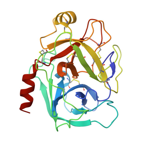
    The magic triangle 5-amino-2,4,6-triiodoisophthalic acid (I3C) and the MAD triangle 5-amino-2,4,6-tribromoisophthalic acid (B3C) are two representatives of a novel class of compounds that combine heavy atoms for experimental phasing with functional groups for protein interactions. These compounds are readily available and provide easy access to experimental phasing. The preparation of stock solutions and the incorporation of the compounds into protein crystals are discussed. As an example of incorporation via cocrystallization, the incorporation of B3C into bovine trypsin, resulting in a single site with high occupancy, is described.


  • Organizational Affiliation

    Department of Structural Chemistry, Georg-August-Universität Göttingen, Tammannstrasse 4, 37077 Göttingen, Germany. tbeck@shelx.uni-ac.gwdg.de


Macromolecules
Find similar proteins by:  (by identity cutoff)  |  3D Structure
Entity ID: 1
MoleculeChains Sequence LengthOrganismDetailsImage
Cationic trypsin223Bos taurusMutation(s): 0 
EC: 3.4.21.4
UniProt
Find proteins for P00760 (Bos taurus)
Explore P00760 
Go to UniProtKB:  P00760
Entity Groups  
Sequence Clusters30% Identity50% Identity70% Identity90% Identity95% Identity100% Identity
UniProt GroupP00760
Sequence Annotations
Expand
  • Reference Sequence
Small Molecules
Binding Affinity Annotations 
IDSourceBinding Affinity
BRV Binding MOAD:  3ITI IC50: 270 (nM) from 1 assay(s)
Experimental Data & Validation

Experimental Data

  • Method: X-RAY DIFFRACTION
  • Resolution: 1.55 Å
  • R-Value Free: 0.213 
  • R-Value Observed: 0.168 
  • Space Group: P 21 21 21
Unit Cell:
Length ( Å )Angle ( ˚ )
a = 53.658α = 90
b = 56.883β = 90
c = 66.808γ = 90
Software Package:
Software NamePurpose
SHELXrefinement
PDB_EXTRACTdata extraction
SAINTdata reduction
SADABSdata scaling
SHELXDphasing
SHELXL-97refinement

Structure Validation

View Full Validation Report


Ligand Structure Quality Assessment 


Entry History 

Deposition Data

Revision History  (Full details and data files)

  • Version 1.0: 2009-10-27
    Type: Initial release
  • Version 1.1: 2011-07-13
    Changes: Version format compliance
  • Version 1.2: 2012-03-21
    Changes: Database references
  • Version 1.3: 2018-01-24
    Changes: Database references