3IOC

Crystal Structure of Mycobacterium Tuberculosis Pantothenate Synthetase at 2.50 Ang resolution in complex with 5'-deoxy-5'-(benzyldisulfanyl)-adenosine


Experimental Data Snapshot

  • Method: X-RAY DIFFRACTION
  • Resolution: 2.50 Å
  • R-Value Free: 0.261 
  • R-Value Work: 0.162 
  • R-Value Observed: 0.166 

wwPDB Validation   3D Report Full Report


Ligand Structure Quality Assessment 


This is version 1.4 of the entry. See complete history


Literature

A Fragment-Based Approach to Probing Adenosine Recognition Sites by Using Dynamic Combinatorial Chemistry

Scott, D.E.Dawes, G.J.Ando, M.Abell, C.Ciulli, A.

(2009) Chembiochem 10: 2772-2779

  • DOI: https://doi.org/10.1002/cbic.200900537
  • Primary Citation of Related Structures:  
    3IOB, 3IOC, 3IOD, 3IOE

  • PubMed Abstract: 

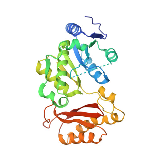
    A new strategy that combines the concepts of fragment-based drug design and dynamic combinatorial chemistry (DCC) for targeting adenosine recognition sites on enzymes is reported. We demonstrate the use of 5'-deoxy-5'-thioadenosine as a noncovalent anchor fragment in dynamic combinatorial libraries templated by Mycobacterium tuberculosis pantothenate synthetase. A benzyl disulfide derivative was identified upon library analysis by HPLC. Structural and binding studies of protein-ligand complexes by X-ray crystallography and isothermal titration calorimetry informed the subsequent optimisation of the DCC hit into a disulfide containing the novel meta-nitrobenzyl fragment that targets the pantoate binding site of pantothenate synthetase. Given the prevalence of adenosine-recognition motifs in enzymes, our results provide a proof-of-concept for using this strategy to probe adjacent pockets for a range of adenosine binding enzymes, including other related adenylate-forming ligases, kinases, and ATPases, as well as NAD(P)(H), CoA and FAD(H2) binding proteins.


  • Organizational Affiliation

    University Chemical Laboratory, University of Cambridge, Lensfield Road, Cambridge, CB2 1EW, UK.


Macromolecules
Find similar proteins by:  (by identity cutoff)  |  3D Structure
Entity ID: 1
MoleculeChains Sequence LengthOrganismDetailsImage
Pantothenate synthetase
A, B
301Mycobacterium tuberculosisMutation(s): 2 
Gene Names: MT3707MTCY07H7B.20panCRv3602c
EC: 6.3.2.1
UniProt
Find proteins for P9WIL5 (Mycobacterium tuberculosis (strain ATCC 25618 / H37Rv))
Explore P9WIL5 
Go to UniProtKB:  P9WIL5
Entity Groups  
Sequence Clusters30% Identity50% Identity70% Identity90% Identity95% Identity100% Identity
UniProt GroupP9WIL5
Sequence Annotations
Expand
  • Reference Sequence
Small Molecules
Binding Affinity Annotations 
IDSourceBinding Affinity
A5D PDBBind:  3IOC Kd: 2.10e+5 (nM) from 1 assay(s)
BindingDB:  3IOC Kd: 2.10e+5 (nM) from 1 assay(s)
ΔH: -5.99e+1 (kJ/mol) from 1 assay(s)
Binding MOAD:  3IOC Kd: 2.10e+5 (nM) from 1 assay(s)
Experimental Data & Validation

Experimental Data

  • Method: X-RAY DIFFRACTION
  • Resolution: 2.50 Å
  • R-Value Free: 0.261 
  • R-Value Work: 0.162 
  • R-Value Observed: 0.166 
  • Space Group: P 1 21 1
Unit Cell:
Length ( Å )Angle ( ˚ )
a = 48.591α = 90
b = 71.485β = 100.03
c = 82.044γ = 90
Software Package:
Software NamePurpose
ADSCdata collection
AMoREphasing
REFMACrefinement

Structure Validation

View Full Validation Report


Ligand Structure Quality Assessment 


Entry History 

Deposition Data

Revision History  (Full details and data files)

  • Version 1.0: 2009-12-01
    Type: Initial release
  • Version 1.1: 2011-07-13
    Changes: Non-polymer description, Version format compliance
  • Version 1.2: 2017-11-01
    Changes: Refinement description
  • Version 1.3: 2021-10-13
    Changes: Database references, Derived calculations
  • Version 1.4: 2023-09-06
    Changes: Data collection, Refinement description