3INT

Structure of UDP-galactopyranose mutase bound to UDP-galactose (reduced)


Experimental Data Snapshot

  • Method: X-RAY DIFFRACTION
  • Resolution: 2.51 Å
  • R-Value Free: 0.247 
  • R-Value Work: 0.194 
  • R-Value Observed: 0.196 

wwPDB Validation   3D Report Full Report


Ligand Structure Quality Assessment 


This is version 1.3 of the entry. See complete history


Literature

X-ray crystallography reveals a reduced substrate complex of UDP-galactopyranose mutase poised for covalent catalysis by flavin .

Gruber, T.D.Westler, W.M.Kiessling, L.L.Forest, K.T.

(2009) Biochemistry 48: 9171-9173

  • DOI: https://doi.org/10.1021/bi901437v
  • Primary Citation of Related Structures:  
    3INR, 3INT

  • PubMed Abstract: 

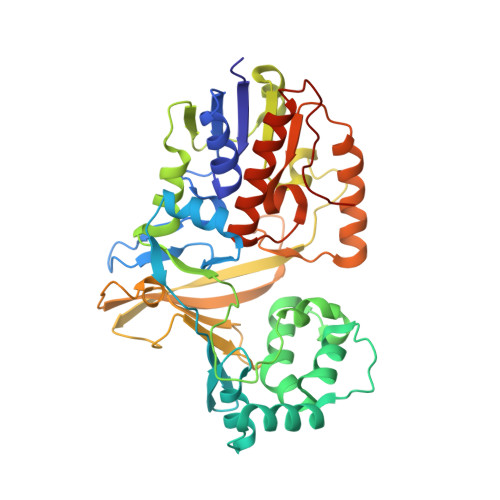
    The flavoenzyme uridine 5'-diphosphate galactopyranose mutase (UGM or Glf) catalyzes the interconversion of UDP-galactopyranose and UDP-galactofuranose. The latter is a key building block for cell wall construction in numerous pathogens, including Mycobacterium tuberculosis. Mechanistic studies of UGM suggested a novel role for the flavin, and we previously provided evidence that the catalytic mechanism proceeds through a covalent flavin-galactose iminium. Here, we describe 2.3 and 2.5 A resolution X-ray crystal structures of the substrate-bound enzyme in oxidized and reduced forms, respectively. In the latter, C1 of the substrate is 3.6 A from the nucleophilic flavin N5 position. This orientation is consistent with covalent catalysis by flavin.


  • Organizational Affiliation

    Department of Biochemistry, National Magnetic Resonance Facility at Madison, University of Wisconsin-Madison, Madison, Wisconsin 53706, USA.


Macromolecules
Find similar proteins by:  (by identity cutoff)  |  3D Structure
Entity ID: 1
MoleculeChains Sequence LengthOrganismDetailsImage
Probable UDP-galactopyranose mutase
A, B
390Klebsiella pneumoniaeMutation(s): 1 
Gene Names: glfrfbD
EC: 5.4.99.9
UniProt
Find proteins for Q48485 (Klebsiella pneumoniae)
Explore Q48485 
Go to UniProtKB:  Q48485
Entity Groups  
Sequence Clusters30% Identity50% Identity70% Identity90% Identity95% Identity100% Identity
UniProt GroupQ48485
Sequence Annotations
Expand
  • Reference Sequence
Experimental Data & Validation

Experimental Data

  • Method: X-RAY DIFFRACTION
  • Resolution: 2.51 Å
  • R-Value Free: 0.247 
  • R-Value Work: 0.194 
  • R-Value Observed: 0.196 
  • Space Group: P 41
Unit Cell:
Length ( Å )Angle ( ˚ )
a = 94.078α = 90
b = 94.078β = 90
c = 129.443γ = 90
Software Package:
Software NamePurpose
HKL-2000data collection
MOLREPphasing
REFMACrefinement
HKL-2000data reduction
HKL-2000data scaling

Structure Validation

View Full Validation Report


Ligand Structure Quality Assessment 


Entry History 

Deposition Data

Revision History  (Full details and data files)

  • Version 1.0: 2009-09-22
    Type: Initial release
  • Version 1.1: 2011-07-13
    Changes: Version format compliance
  • Version 1.2: 2021-10-13
    Changes: Database references, Derived calculations
  • Version 1.3: 2023-09-06
    Changes: Data collection, Refinement description