3I68

Plasmodium falciparum dihydroorotate dehydrogenase bound with triazolopyrimidine-based inhibitor DSM2


Experimental Data Snapshot

  • Method: X-RAY DIFFRACTION
  • Resolution: 2.40 Å
  • R-Value Free: 0.267 
  • R-Value Work: 0.214 
  • R-Value Observed: 0.214 

wwPDB Validation   3D Report Full Report


Ligand Structure Quality Assessment 


This is version 1.2 of the entry. See complete history


Literature

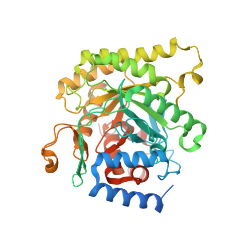
Structural plasticity of malaria dihydroorotate dehydrogenase allows selective binding of diverse chemical scaffolds.

Deng, X.Gujjar, R.El Mazouni, F.Kaminsky, W.Malmquist, N.A.Goldsmith, E.J.Rathod, P.K.Phillips, M.A.

(2009) J Biol Chem 284: 26999-27009

  • DOI: https://doi.org/10.1074/jbc.M109.028589
  • Primary Citation of Related Structures:  
    3I65, 3I68, 3I6R

  • PubMed Abstract: 

    Malaria remains a major global health burden and current drug therapies are compromised by resistance. Plasmodium falciparum dihydroorotate dehydrogenase (PfDHODH) was validated as a new drug target through the identification of potent and selective triazolopyrimidine-based DHODH inhibitors with anti-malarial activity in vivo. Here we report x-ray structure determination of PfDHODH bound to three inhibitors from this series, representing the first of the enzyme bound to malaria specific inhibitors. We demonstrate that conformational flexibility results in an unexpected binding mode identifying a new hydrophobic pocket on the enzyme. Importantly this plasticity allows PfDHODH to bind inhibitors from different chemical classes and to accommodate inhibitor modifications during lead optimization, increasing the value of PfDHODH as a drug target. A second discovery, based on small molecule crystallography, is that the triazolopyrimidines populate a resonance form that promotes charge separation. These intrinsic dipoles allow formation of energetically favorable H-bond interactions with the enzyme. The importance of delocalization to binding affinity was supported by site-directed mutagenesis and the demonstration that triazolopyrimidine analogs that lack this intrinsic dipole are inactive. Finally, the PfDHODH-triazolopyrimidine bound structures provide considerable new insight into species-selective inhibitor binding in this enzyme family. Together, these studies will directly impact efforts to exploit PfDHODH for the development of anti-malarial chemotherapy.


  • Organizational Affiliation

    Department of Pharmacology, University of Texas Southwestern Medical Center at Dallas, Dallas, Texas 75390-9041, USA.


Macromolecules
Find similar proteins by:  (by identity cutoff)  |  3D Structure
Entity ID: 1
MoleculeChains Sequence LengthOrganismDetailsImage
Dihydroorotate dehydrogenase homolog, mitochondrial415Plasmodium falciparum 3D7Mutation(s): 0 
Gene Names: PFF0160c
EC: 1.3.3.1
Membrane Entity: Yes 
UniProt
Find proteins for Q08210 (Plasmodium falciparum (isolate 3D7))
Explore Q08210 
Go to UniProtKB:  Q08210
Entity Groups  
Sequence Clusters30% Identity50% Identity70% Identity90% Identity95% Identity100% Identity
UniProt GroupQ08210
Sequence Annotations
Expand
  • Reference Sequence
Small Molecules
Ligands 3 Unique
IDChains Name / Formula / InChI Key2D Diagram3D Interactions
FMN
Query on FMN

Download Ideal Coordinates CCD File 
C [auth A]FLAVIN MONONUCLEOTIDE
C17 H21 N4 O9 P
FVTCRASFADXXNN-SCRDCRAPSA-N
J4Z
Query on J4Z

Download Ideal Coordinates CCD File 
B [auth A]N-anthracen-2-yl-5-methyl[1,2,4]triazolo[1,5-a]pyrimidin-7-amine
C20 H15 N5
OXYDLVAOXASTMW-UHFFFAOYSA-N
ORO
Query on ORO

Download Ideal Coordinates CCD File 
D [auth A]OROTIC ACID
C5 H4 N2 O4
PXQPEWDEAKTCGB-UHFFFAOYSA-N
Binding Affinity Annotations 
IDSourceBinding Affinity
J4Z BindingDB:  3I68 IC50: 56 (nM) from 1 assay(s)
Binding MOAD:  3I68 IC50: 56 (nM) from 1 assay(s)
Experimental Data & Validation

Experimental Data

  • Method: X-RAY DIFFRACTION
  • Resolution: 2.40 Å
  • R-Value Free: 0.267 
  • R-Value Work: 0.214 
  • R-Value Observed: 0.214 
  • Space Group: P 64
Unit Cell:
Length ( Å )Angle ( ˚ )
a = 85.917α = 90
b = 85.917β = 90
c = 138.697γ = 120
Software Package:
Software NamePurpose
HKL-2000data collection
PHASERphasing
REFMACrefinement
HKL-2000data reduction
HKL-2000data scaling

Structure Validation

View Full Validation Report


Ligand Structure Quality Assessment 


Entry History 

Deposition Data

Revision History  (Full details and data files)

  • Version 1.0: 2009-07-28
    Type: Initial release
  • Version 1.1: 2011-07-13
    Changes: Version format compliance
  • Version 1.2: 2023-09-06
    Changes: Data collection, Database references, Derived calculations, Refinement description