3HUS

Crystal structure of recombinant gamma N308K fibrinogen fragment D with the peptide ligand Gly-Pro-Arg-Pro-amide


Experimental Data Snapshot

  • Method: X-RAY DIFFRACTION
  • Resolution: 3.04 Å
  • R-Value Free: 0.288 
  • R-Value Work: 0.220 
  • R-Value Observed: 0.238 

wwPDB Validation   3D Report Full Report


This is version 2.2 of the entry. See complete history


Literature

Impaired protofibril formation in fibrinogen gammaN308K is due to altered D:D and "A:a" interactions.

Bowley, S.R.Okumura, N.Lord, S.T.

(2009) Biochemistry 48: 8656-8663

  • DOI: https://doi.org/10.1021/bi900239b
  • Primary Citation of Related Structures:  
    3HUS

  • PubMed Abstract: 

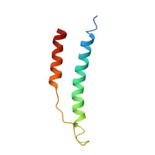
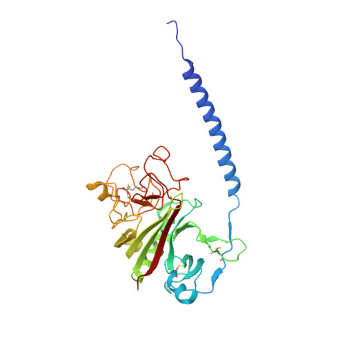
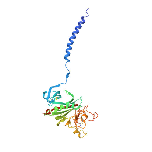
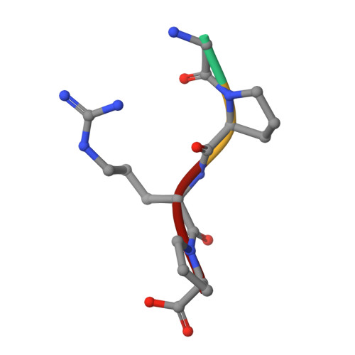
    "A:a" knob-hole interactions and D:D interfacial interactions are important for fibrin polymerization. Previous studies with recombinant gammaN308K fibrinogen, a substitution at the D:D interface, showed impaired polymerization. We examined the molecular basis for this loss of function by solving the crystal structure of gammaN308K fragment D. In contrast to previous fragment D crystals, the gammaN308K crystals belonged to a tetragonal space group with an unusually long unit cell (a = b = 95 A, c = 448.3 A). Alignment of the normal and gammaN308K structures showed the global structure of the variant was not changed and the knob "A" peptide GPRP was bound as usual to hole "a". The substitution introduced an elongated positively charged patch in the D:D region. The structure showed novel, symmetric D:D crystal contacts between gammaN308K molecules, indicating the normal asymmetric D:D interface in fibrin would be unstable in this variant. We examined GPRP binding to gammaN308K in solution by plasmin protection assay. The results showed weaker peptide binding, suggesting that "A:a" interactions were altered. We examined fibrin network structures by scanning electron microscopy and found the variant fibers were thicker and more heterogeneous than normal fibers. Considered together, our structural and biochemical studies indicate both "A:a" and D:D interactions are weaker. We conclude that stable protofibrils cannot assemble from gammaN308K monomers, leading to impaired polymerization.


  • Organizational Affiliation

    Department of Chemistry, University of North Carolina at Chapel Hill, Chapel Hill, North Carolina 27599, USA.


Macromolecules
Find similar proteins by:  (by identity cutoff)  |  3D Structure
Entity ID: 1
MoleculeChains Sequence LengthOrganismDetailsImage
Fibrinogen alpha chain
A, D
66Homo sapiensMutation(s): 0 
Gene Names: FGA
UniProt & NIH Common Fund Data Resources
Find proteins for P02671 (Homo sapiens)
Explore P02671 
Go to UniProtKB:  P02671
PHAROS:  P02671
GTEx:  ENSG00000171560 
Entity Groups  
Sequence Clusters30% Identity50% Identity70% Identity90% Identity95% Identity100% Identity
UniProt GroupP02671
Sequence Annotations
Expand
  • Reference Sequence
Find similar proteins by:  (by identity cutoff)  |  3D Structure
Entity ID: 2
MoleculeChains Sequence LengthOrganismDetailsImage
Fibrinogen beta chain
B, E
313Homo sapiensMutation(s): 0 
Gene Names: FGB
UniProt & NIH Common Fund Data Resources
Find proteins for P02675 (Homo sapiens)
Explore P02675 
Go to UniProtKB:  P02675
PHAROS:  P02675
GTEx:  ENSG00000171564 
Entity Groups  
Sequence Clusters30% Identity50% Identity70% Identity90% Identity95% Identity100% Identity
UniProt GroupP02675
Sequence Annotations
Expand
  • Reference Sequence
Find similar proteins by:  (by identity cutoff)  |  3D Structure
Entity ID: 3
MoleculeChains Sequence LengthOrganismDetailsImage
Fibrinogen gamma chain
C, F
311Homo sapiensMutation(s): 1 
Gene Names: FGGPRO2061
UniProt & NIH Common Fund Data Resources
Find proteins for P02679 (Homo sapiens)
Explore P02679 
Go to UniProtKB:  P02679
PHAROS:  P02679
GTEx:  ENSG00000171557 
Entity Groups  
Sequence Clusters30% Identity50% Identity70% Identity90% Identity95% Identity100% Identity
UniProt GroupP02679
Sequence Annotations
Expand
  • Reference Sequence

Find similar proteins by:  Sequence   |   3D Structure  

Entity ID: 4
MoleculeChains Sequence LengthOrganismDetailsImage
Peptide Ligand Gly-Pro-Arg-Pro-amide
G, H, I, J
4N/AMutation(s): 0 
Sequence Annotations
Expand
  • Reference Sequence
Oligosaccharides

Help

Entity ID: 5
MoleculeChains Length2D Diagram Glycosylation3D Interactions
2-acetamido-2-deoxy-beta-D-glucopyranose-(1-4)-[alpha-L-fucopyranose-(1-6)]2-acetamido-2-deoxy-beta-D-glucopyranose
K, L
3N-Glycosylation
Glycosylation Resources
GlyTouCan:  G21290RB
GlyCosmos:  G21290RB
GlyGen:  G21290RB
Experimental Data & Validation

Experimental Data

  • Method: X-RAY DIFFRACTION
  • Resolution: 3.04 Å
  • R-Value Free: 0.288 
  • R-Value Work: 0.220 
  • R-Value Observed: 0.238 
  • Space Group: P 43 21 2
Unit Cell:
Length ( Å )Angle ( ˚ )
a = 95.009α = 90
b = 95.009β = 90
c = 448.382γ = 90
Software Package:
Software NamePurpose
HKL-2000data collection
PHASERphasing
REFMACrefinement
HKL-2000data reduction
HKL-2000data scaling

Structure Validation

View Full Validation Report


Entry History 

Deposition Data

Revision History  (Full details and data files)

  • Version 1.0: 2009-08-18
    Type: Initial release
  • Version 1.1: 2011-07-13
    Changes: Advisory, Version format compliance
  • Version 2.0: 2020-07-29
    Type: Remediation
    Reason: Carbohydrate remediation
    Changes: Advisory, Atomic model, Data collection, Derived calculations, Structure summary
  • Version 2.1: 2021-10-13
    Changes: Database references, Structure summary
  • Version 2.2: 2023-09-06
    Changes: Data collection, Refinement description